Bronchial Malignant Melanoma
Key Facts
Clinical Issues
Incidence
Approximately 0.01% of all lung neoplasms
More common in adults in 6th decade of life
Symptoms
Cough
Dyspnea
Chest pain hemoptysis
Fever
Endoscopic findings
Possible pigmented lesion
Microscopic Pathology
Nested pattern
Spindle cell pattern
Pigmented lesion
Top Differential Diagnoses
Carcinoma
Will show negative staining for S100 protein, HMB-45, &/or mart-1
Neuroendocrine carcinoma
Melanomas are generally negative for neuroendocrine markers, i.e., chromogranin-A and synaptophysin
Neurogenic sarcoma
S100 may show positive staining in both tumors
Negative for HMB-45 and mart-1
Metastatic melanoma
Clinical history is most important and reliable way to determine primary site
Ocular evaluation is highly important
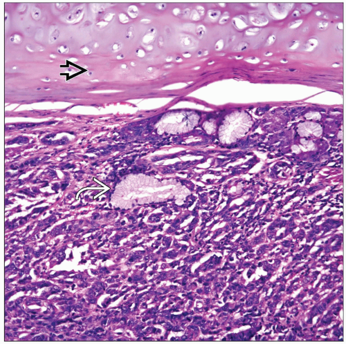 Primary bronchial melanoma involving full thickness of the bronchial epithelium. Note the presence of residual endobronchial glandular structures  as well as bronchial cartilage as well as bronchial cartilage  . . |
 High-power view of a bronchial malignant melanoma shows prominent nuclear atypia and mitotic activity. Cellular pleomorphism is a common feature of bronchial melanoma |
TERMINOLOGY
Synonyms
Pulmonary malignant melanoma
Definitions
Malignant neoplasm of possible neural crest derivation
ETIOLOGY/PATHOGENESIS
Etiology
No specific etiology for these tumors when they occur in the lung
CLINICAL ISSUES
Epidemiology
Incidence
Estimated that primary pulmonary melanomas represent approximately 0.01% of all lung neoplasms
Age
More common in adults in 6th decade of life
Gender
No gender predilection
Presentation
Cough
Shortness of breath
Chest pain
Hemoptysis
Fever
Endoscopic Findings
Possible pigmented lesion
Treatment
Surgical approaches
Lobectomy or pneumonectomy
Adjuvant therapy
Probable chemotherapy
Prognosis
Variable
Death within 30 months after initial diagnosis
Possible survival at 5 years of 40%
MACROSCOPIC FEATURES
General Features
Well-defined tumor mass
Pigment
Homogeneous surface
Areas of necrosis &/or hemorrhage may be seen
Polypoid bronchial mass in some cases
MICROSCOPIC PATHOLOGY
Histologic Features
Nested pattern
Spindle cell pattern
Pigmented lesion
Cells with prominent nucleoli
Stay updated, free articles. Join our Telegram channel

Full access? Get Clinical Tree


