4 Breast Diseases
Anatomy of the Breast
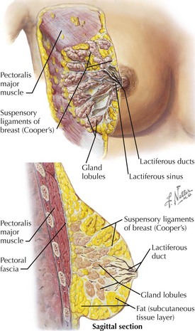
Basic Structure
• Adipose tissue and lactiferous glands lie between superficial and deep layers of superficial thoracic fascia.
• Cooper’s (suspensory) ligaments: partitions of fibrous connective tissue running from the deep fascia over the pectoralis major, external intercostals, and serratus anterior, through the breast parenchyma, to the dermis and superficial fascia
Endocrinology
• Estrogen (e.g., in pregnancy) and tissue-based estrogen receptors control glandular proliferation and secretory states in concert with progesterone and other hormones and growth factors.
Vessels and Lymphatics
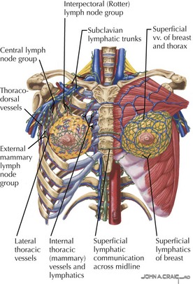
Arterial Supply
• Lateral: thoracoacromial, lateral thoracic, circumflex scapular, subscapular, and thoracodorsal branches of the axillary artery (second and third divisions)
Venous Drainage
• Drainage toward the axilla and axillary vein via named branches, including supreme thoracic, thoracoacromial, lateral thoracic, circumflex scapular, subscapular, and thoracodorsal


Lymphatic Drainage


• Deep flow is along internal thoracic (mammary) vessel pathways to parasternal nodes, draining toward subclavian, supraclavicular, and deep cervical nodes.
• Location of sentinel node (nearest, with metastasis) in axilla depends on the patient’s specific drainage pattern from the tumor site.


