Bone Manifestations of Hematologic Disorders and Small Cell Tumors


Bone Manifestations of Hematologic Disorders and Small Cell Tumors


Jeffrey F. Lipton, M.D

Vincent J. Vigorita, M.D.


The osseous and articular changes of hematologic disorders consist of primary or secondary effects on bone and joint tissues.

The extent of bone change is directly related to the age of the patient at onset and the severity of associated hematopoietic tissue alteration, especially the erythroid hyperplasia that accompanies all hemolytic anemias. Because hematopoietic bone marrow in infants is present in the bones of the hands and feet as well as other bones, the osseous changes of hematologic disorders in the first few years of life may be dramatic at these sites (1) (Fig. 12.1). In childhood, red marrow is more in evidence in the bones of the skull. Final anatomic locations of hematopoietic tissue are the vertebral column, sternum, ribs, and pelvis.

Red marrow develops during fetal life in all bones and in the liver and spleen (Fig. 12.2). At birth, extramedullary hematopoiesis ceases, and red marrow begins to taper off. Shortly after skeletal maturation is achieved, the long bones contain essentially fatty marrow. Red marrow at the other sites tapers off gradually until midlife, plateaus during the middle years, and tapers off thereafter. Aging bones increasingly contain fatty marrow, which may be related to two important age-related disorders: anemia and osteoporosis.


Hemolytic Anemia


Sickle Cell Anemia

Hemoglobin, the oxygen-carrying moiety of blood, consists of four heme-globin units, each containing a disc-shaped molecule with iron in the center. Surrounding each heme (identical in each hemoglobin molecule) are two types of globin peptide chains. Normal adult blood consists of several hemoglobins (hemoglobin A, 90 percent; hemoglobin A2, 2.5 percent; hemoglobin F, less than 1 percent) (Table 12.1). There are no other normal hemoglobin variants in adult blood. Hemoglobin A contains two α chains and two β chains. In other hemoglobins, the α chains are the same. The companion chains are different in all other hemoglobins.

Hemoglobin F has two γ chains, and hemoglobin A2 has two δ chains. Although hemoglobin A2 is not clinically significant per se, elevated levels are seen in β-thalassemia trait. Hemoglobin F is actually therapeutic in sickle cell anemia, and modern therapies are in part directed at increasing its level with substances like hydroxyurea (2,3). Experimentally and clinically, hemoglobin F inhibits the damaging polymerization that sickle cell hemoglobin undergoes.

Other hemoglobin variants are usually the result of genetically determined substitutions of amino acids in one of the globin chains, usually an alteration in the β chain. Sickle cell anemia is caused by formation of the hemoglobin variant S, in which valine becomes the sixth amino acid in the β chain, replacing glutamic acid. Hemoglobin S quantitatively replaces hemoglobin A. Whether an individual has sickle cell trait or sickle cell disease depends on the percentages of hemoglobins A, A2, F, and S present (Table 12.2).

The most common method of identifying hemoglobin variants, such as sickle cell hemoglobin S, is to place hemolysate on a cellulose acetate strip (pH 8.4) and subject it to a voltage drop. Hemoglobins migrate depending on different charges. Further electrophoresis plating (on citrate agar) differentiates hemoglobin S. New methods use monoclonal antibodies to detect the presence of hemoglobin S.

Hemoglobin S is susceptible to deoxygenation. It is relatively insoluble and polymerizes. The dangerous polymerization of hemoglobin S increases progressively as oxygen saturation (of whatever cause) decreases. Rigid, physiologically compromising shapes result from hemoglobin S polymerization, impairing microcirculation (Fig. 12.3).

The end result includes breakdown of red blood cells (chronic hemolytic anemia), progressive tissue damage, and painful acute
vaso-occlusive crises. Infarction results from vaso-occlusive events (Fig. 12.4). The rigid sickle cells become sequestered by the reticuloendothelial system (spleen), which leads to shortened red cell survival. Massive blood pooling in the spleen can occur.


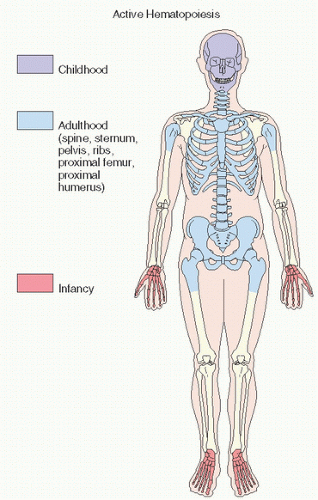
FIGURE 12.1. Sites of active hematopoiesis at different stages of life.


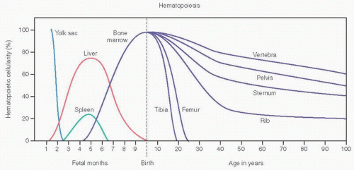
FIGURE 12.2. Hematopoiesis and age. (Adapted from Lukens JN. Blood formation in the embryo, fetus, and newborn. In: Lee CR, et al., eds. Wintrobe’s Clinical Hematology. 9th ed. Philadelphia, PA: Lea & Febiger; 1993:79-100.)


TABLE 12.1 Normal Hemoglobins (Hb) in Adults


Hb


Concentration (%)


Structure


A


≈90


α2β2


F


<1


α2γ2


A2


1.5-≈2.5


α2δ2


Crises can be precipitated or worsened by infection, dehydration, acidosis, and hypoxia of whatever etiology.

Approximately 150 black children in 100,000 have sickle cell anemia, inheriting hemoglobin S from both parents. About 1 in 12 has the trait, inheriting hemoglobin S from only one parent. In basic mendelian genetics, a child born of two parents, each carrying the trait, has a 50 percent chance of developing sickle cell trait, a 25 percent chance of developing sickle cell anemia, and a 25 percent chance of being normal. Roughly 10 percent of blacks in America have the sickle cell trait.

Periodic, self-limited attacks of severe musculoskeletal pain characterize the lives of sickle cell patients (4). Platt et al. (4) estimated 0.8 episodes of pain per patient-year. These so-called crises cause considerable morbidity, and their frequency has been correlated with early death in patients older than the age of 20 years. The pain, which has been likened to that of a severe toothache, affects multiple sites including extremities, lower back, and abdomen.

In addition to pain, numerous orthopaedic bone and joint complications have been documented in sicklers. Most of the musculoskeletal problems associated with sickle cell disease are the result of vaso-occlusive complications including avascular necrosis (5), osteomyelitis, septic arthritis, myonecrosis, reactive arthritis, and dactylitis (6). In addition, slipped capital femoral epiphysis and
growth disturbances can be seen. In one study of 57 sicklers from Saudi Arabia, Bennett and Namnyak (7) detailed the clinical problems (Table 12.3). The hand and foot syndrome, an early manifestation in about one-third of cases, is thought to be a manifestation of bone and marrow infarction.


TABLE 12.2 Percentages of Hemoglobin (Hb) in Normal Persons and Those with Sickle Cell Disease and Sickle Cell Trait


HbA


HbS


HbF


HbA2


Normal


˜95%


0


<1%


1.5%-3.5%


Sickle cell disease (also HbD, HbG)


0


>80%


1%-20%


2%-4.5%


Sickle cell trait


50%-60%


34%-45%


<1%


<4.5%


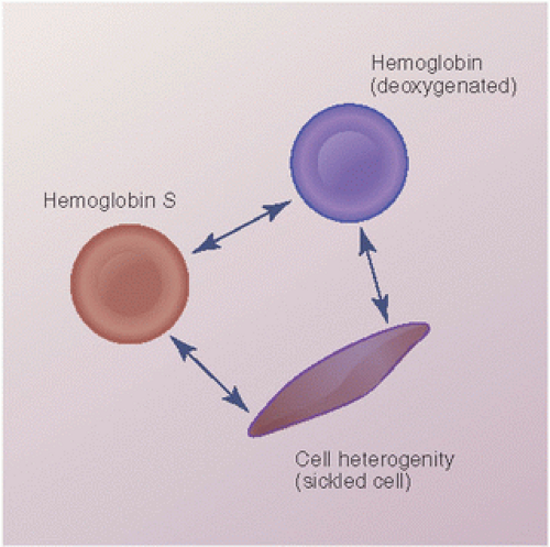
FIGURE 12.3. The progression of hemoglobin S to a deoxygenated state, and its further polymerized state seen as a sickled cell.

Nonorthopaedic medical problems include congestive heart failure, splenic crisis, chest syndrome, right upper quadrant syndrome, retinopathy, and pulmonary hypertension.

In sickle cell disease, roentgenographic changes can be attributed to secondary erythroid hyperplasia, ischemic bone changes, and osteomyelitis. Erythroid hyperplasia, similar to thalassemia, leads to thin cortices and medullary widening, manifested as a tower-shaped skull, kyphosis, scoliosis, and saber shins. Vertebral cupping (anteriorly) and “hair on end” skull appearances are rare but well documented.

The most common radiographic findings for sickle cell anemia include osteoporosis, endosteal apposition of bone, infarctions, and infections. The osteoporosis can be diffuse, present throughout the skeleton. The endosteal formation of new bone often results in a “bone within a bone” appearance. Areas of infarction can occur at the ends of bones, such as the femoral head, and in the shafts of bones. The differential diagnosis in these situations includes Gaucher disease, Legg-Calvé-Perthes disease, and any other causes of bone infarctions.

Infection can be superimposed on sickle cell anemia, and can be difficult to differentiate from bone infarction (8). In both conditions, the child may have a fever and a swollen, painful, and tender leg with limited range of motion. A combination of sequential bone marrow scan (0.280 m Ci/kg of technetium-99m sulfur colloid) and bone scintigraphy (0.289 m Ci/kg technetium methylene diphosphonate) may be useful: Infarction is most probable with a reduced activity on bone marrow scan and abnormal activity on bone scan. In contrast, osteomyelitis is more probable with a normal bone marrow scan and abnormal bone scan (8). The usual radiographic manifestations of osteomyelitis are present in sickle cell disease: areas of lucency, sometimes with a dense sequestrum and surrounded by sclerosis in chronic cases, and new periosteal bone reaction in acute cases.

Frontal and lateral views of the spine often demonstrate a collapse of the central portion of the vertebral end plate, thought to be secondary to an anatomically poor vascular supply to this portion of the end plate. Further compromise of the blood supply, such as occurs in a sickler, affects the central end plate before other portions of the vertebra.

In infants, periosteal elevations in metacarpal and metatarsal bones are seen, as well as shortening of the first metacarpal. Severe periosteal reactions in the large tubular bones in early childhood and a hair-on-end appearance and thickened skull in late childhood are seen. In adult sicklers, the ends of long bones near weight-bearing surfaces are most affected. Initially lytic, they become sclerotic as well. Osteonecrosis with cortical thickening is thought to be typical of sickle cell disease.

Osteonecrosis of the femoral head is particularly common in sickle cell disease and has been conservatively estimated to occur in at least 10 percent of patients. Results of hip arthroplasty are poor (8,9). Osteomyelitis is a major skeletal complication, and, as mentioned, can be extremely difficult to distinguish from acute bone infarction (10).

Sicklers are prone to Salmonella osteomyelitis, and Salmonella infection has been shown to be the predominant cause of osteomyelitis in sicklers in African and Saudi Arabian studies (11,12,13). American studies show a predominance of Staphylococcus aureus in sicklers (9). Septic Salmonella arthritis is well described (14) (Fig. 12.5).

Skin ulcers extending deep into soft tissues and bone may lead to radiographic changes, including periosteal reactions and osteoma-like cortical remodeling (15).

Few biologic benefits derive from sickle cell disease, but decreased susceptibility to malaria is one. It is most convincing for sickle trait, and most probably is related to inhibition of the growth of Plasmodium falciparum in sickle erythrocytes, attributable to sequestration of infected red blood cells along venules with low oxygen tension or the development of a tolerance mechanism.

High levels of fetal hemoglobin can predict improved survival and have been proposed as a reliable childhood indicator of adult life expectancy (16).


FIGURE 12.4. Peripheral smears from a normal person (A), a person with iron deficiency (B), a patient with thalassemia (C), and a sickler (D). Normal red blood cells have a slightly ovoid shape with central pallor. In iron deficiency and thalassemia, cells are small (microcytic) and pale and have less hemoglobin (hypochromic). In more severe thalassemia (C), hemolytic changes become apparent, including the presence of target cells and broken, fragmented cells. In (D), the classic crescent-shaped cells of a sickler are evident.

No single treatment is effective for all patients with sickle cell disease. Avoiding dehydration, extreme temperatures, and conditions of low oxygen levels such as high altitudes and intense physical training are important. Hydroxyurea has been the backbone of management as it induces fetal hemoglobin production, which possesses a strong affinity for oxygen binding.


TABLE 12.3 Clinical Details in 57 Patients with Sickle Cell Anemia


Initial Findings


No. of Patients


Temperature <37 °C


47


Jaundice


5


Splenomegaly


19


Cellulitis


7


Bone and joint pain


46


Deep soft tissue abscess


8


Flexion-adduction contracture of the hip


3


Skin ulceration


2


Septic arthritis


4


Osteomyelitis


35


Joint effusion


8


Hemarthrosis


2


Modified after Bennett OM, Namnyak SS. Bone and joint manifestations of sickle cell anaemia. J Bone Joint Surg Br. 1990;72:494-499.


Transplantation is the only known cure and involves blood or bone marrow stem cell transplantation.


Thalassemia (Cooley Anemia)

Thalassemia is a common hemolytic anemia that is a consequence of abnormal hemoglobin synthesis. Among the most common genetic disorders worldwide, it has been estimated at 0.44 per 1,000 births (17). In β-thalassemia, there is a relative decrease or absence of the β chain (hemoglobin normally consisting of a tetramer of two α-chains and two β chains) with a resultant excess of α-chains. Normal red blood cells contain a mixture of hemoglobins, comprising approximately 90 percent hemoglobin A, 2 percent hemoglobin A2, and 1 percent fetal hemoglobin (Table 12.1). In thalassemia, the proportions are altered (Table 12.4). It is a relatively common disorder of people of Mediterranean origin. Red blood cells become hypochromic and microcytic, as in iron-deficiency anemia (Fig. 12.4). However, cells break down, causing hemolytic changes visualized as precipitated hemoglobin moieties in target cells identifiable on peripheral blood smears. The red marrow responds to compensate for the ineffective erythropoiesis.


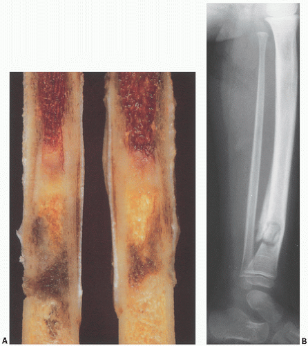
FIGURE 12.5. (A) Sickle cell anemia. Amputated tibia shows viable hyperplastic red marrow proximally and gradations of infarction distally. (B) Osteomyelitis in a case of sickle cell anemia. Areas of sclerosis and lucency in the tibia are surrounded by periosteal reaction. Pathologic fracture is present. The infection was caused by Staphylococcus aureus.

Bone changes reflect the severity of disease and are therefore less profound in thalassemia minor. It is a prototype disease for studying the osseous effects of hemolytic anemia (1).

In general, there is less involvement of the peripheral skeleton as the patient matures, although severe thalassemia present in infancy has profound, roentgenographically detectable effects (1,18,19).

The radiographic findings reflect marrow hyperactivity as the marrow attempts to compensate for the hemolytic anemia (Fig. 12.6) (18). The findings are most prominent in the major form of the disease and can be completely absent in the minor form. The major form usually manifests itself in the first 2 years of life. These children tend to be short in stature, the development of secondary sexual characteristics is usually delayed, and the facies tend to be mongoloid or rodent-like in appearance.


TABLE 12.4 Characteristics of β-Thalassemias


Condition


Parental Genotypes


Risk


Hemoglobin Patterna


Severity


β mRNA


Genes


Homozygous states


Both β+


1/4


↓HbA, ↑HbF, variable HbA2


Variable, usually Cooley’s anemia


Marked deficiency of β mRNA


β genes present


Heterozygous states


β+/β, normal


1/2


↑HbA2, slight ↑HbF


Thalassemia minor


Deficient β mRNA


β genes present


a Normal: 90% A1, 2% A2, 1% F.


β+ = thalassemia


The hyperplasia of the bone marrow (often five to six times normal cellularity) leads to the destruction of medullary bone trabeculae, expansion of the bone, and cortical thinning. These changes can occur throughout the skeleton; however, they tend to be more prominent in the hands and feet. The short tubular bones are more lucent than usual. These bones are expanded, and the remaining trabeculae are accentuated. Abnormal modeling of the


bones is found in thalassemia major. This is most commonly seen in the distal femur, where hyperactive marrow causes widening of the metaphyses and “Erlenmeyer flask” deformity, which can also be found in Gaucher disease, Pyle disease, and osteopetrosis. Similar metaphyseal deformity can be seen in other long bones of patients with these diseases. A “cobwebbing” pattern may be seen in trabecular bone.


FIGURE 12.6. (A) The pathogenesis of bone and bone marrow changes in β-thalassemia resulting from hemolysis and ineffective erythropoiesis. (Modified from Olivieri, NF. The β-thalassemias. N Engl J Med. 1999;341:99-109.) (B) Thalassemia. Changes in the hand (a) and upper extremity (Continued)


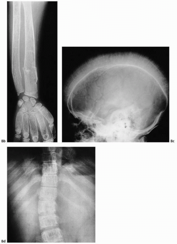
FIGURE 12.6. (Continued) (b) include medullary widening, cortical thinning, marked osteoporosis, and accentuation of the trabeculae of all short tubular bones. The skull may show a hair-on-end appearance (c). In (d), accentuation of trabeculae of the spine and widening of posterior portions of the rib are seen.

In the skull, thalassemia major often causes widening of the diploic cavity in most of the calvaria, with relative sparing of the occiput, where marrow is not as prominent as in the rest of the skull. Thin, perpendicular bone spicules result at times in a hair-on-end or “hairbrush” appearance. The paranasal sinuses are poorly formed or not developed at all in the most severe forms of the disease. This is the result of encroachment on the region of the paranasal sinuses by soft tissue masses, areas of extramedullary hematopoiesis. The term rodent facies has been used to describe the end result of facial changes.

Extramedullary hematopoiesis can also occur in soft tissue masses in the chest or abdomen (20). These masses are most common close to the spine and are, at times, associated with expansion and cortical thinning of the adjacent vertebrae and ribs. Correction of the underlying anemia often results in shrinking or disappearance of these soft tissue masses.

Treating the hemolytic anemia of thalassemia has required repeated blood transfusions, which introduces one of the severe complications of the disease: secondary hemosiderosis from iron accumulation resulting from transfusion overload. Iron deposition is demonstrable in both bone and synovium, iron overload causing most of the mortality and morbidity associated with thalassemia (17). It has been estimated that a minimum of 100 transfusions is required to cause iron-related disease (hemochromatosis) (21). In bone, iron localizes at the mineralization front (Fig. 12.7). Only recently, however, has bone disease been identified. Osteoporosis and rare cases of osteomalacia have been documented.

In the joint, a specific migratory polyarthritis dominates the clinical picture (20). Aseptic necrosis of bone, proposed to be caused by chronic anemic hypoxia, has been reported.

Treatment is directed at chelating the iron of transfusion hemosiderosis. Both intravenous (deferoxamine) and oral (deferiprone) chelating agents have been used, as well as hydroxy urea. Although the most common iron-chelating agent used, deferoxamine has several limitations including the need for parenteral administration (and its associated pain and reduced compliance), cost, and side effects (17). Exogenous hepcidin or hepcidin-signaling agonists are being explored.


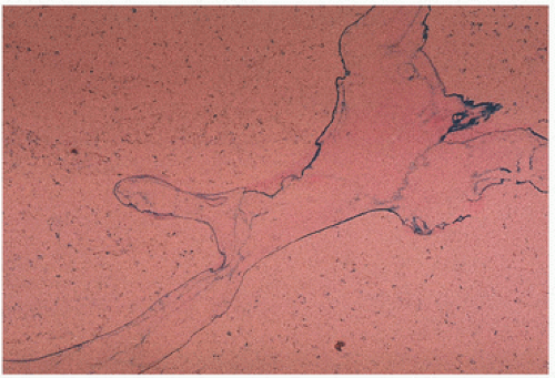
FIGURE 12.7. Thalassemia. Iron (blue) is localized to the mineralizing fronts of bone (Prussian blue stain).


Hemophilia

Hemophilia A, the most common hereditary coagulation disorder, affects 20 live male infants per 100,000. It is caused by the deficiency, absence, or malfunction of coagulation factor VIII (22). In normal patients, a very low concentration of factor VIII (0.2 µ/mL plasma) is sufficient for adequate coagulation. Bleeding leading to clinically perceptible illness requires a reduction of at least 75 percent.

Hemophilia A is a clinically heterogeneous disorder, ranging from mild (1 to 4 percent deficiency) to severe disease. Patients with mild or moderate disease may not be recognized unless a significant traumatic event precipitates abnormal bleeding. Excessive bleeding during surgery may be the first clue. Therapy centers around replacement of factor VIII, which was traditionally done with shotgun frozen plasma. Cryoprecipitate became available in the 1960s. More recently, purified concentrates have been used. In general, 1 U of factor VIII increases plasma activity by 0.024/mL. Because 0.3 U/mL is usually needed to treat a mild episode of bleeding, clinical goals should strive for more than this amount. Therapy has significantly decreased but not eliminated morbidity from the disorder (22).

Clinically, abnormal hemorrhaging is key, especially into joints. Most commonly involved, in order of frequency, are the knees and elbows, followed by the ankles, shoulders, and hips. Not usually evident in infancy, childhood signs include mild discomfort and limitation of joint motion. Pain and swelling follow. Numerous damaging microhemarthroses have transpired before clinical suspicion is initially aroused, so that the diagnosis is unfortunately delayed.

The pathology centers around iron-induced synovial inflammation (see Fig. 15.12), with both clinical and experimental observations documenting the damaging effects of hemosiderosis synovitis.

Roentgenographically, narrowing of joint spaces, loss of articular cartilage, cystic remodeling of bone (Fig. 12.8), and hemophilic pseudotumors characterize the illness. However, scrupulous adherence to maintenance of factor VIII levels to prevent spontaneous hemorrhage has been associated with significantly decreased morbidity and has diminished but not eliminated the radiographic progression of disease.

Involvement of the musculoskeletal system is one of the major complications of hemophilia. The knee is the most commonly involved joint, followed by the elbow, ankle, shoulder, and hip. Hemophiliac arthropathy in the small joints of the hands and feet is rare, with few series of cases described in the literature.

The radiographic presentation of hemophiliac arthropathy can be divided into stages for didactic purposes, but in real practice, there is frequent overlap between the different phases of the disease. Stage I is acute bleeding in the joint. Distension of the articular capsule is easily seen as a distended suprapatellar bursa in the case of the knee, or displacement of the humeral fat pads in the case of the elbow. Distension of the ankle joint capsule is usually easy to detect in the lateral projection. Bleeding into the shoulder or hip joints is difficult to determine in plain radiographs, but can be assessed by magnetic resonance imaging (MRI) and, to a lesser degree, also by computed tomography (CT).


FIGURE 12.8. Cystic remodeling of the glenohumeral joint in a patient with hemophilia. (A) Large subchondral erosions are present in the humeral head and glenoid. (B) Computed tomography (CT) shows areas of increased density in the soft tissues, secondary to hemosiderin deposition in the capsule of the joint. (Continued)


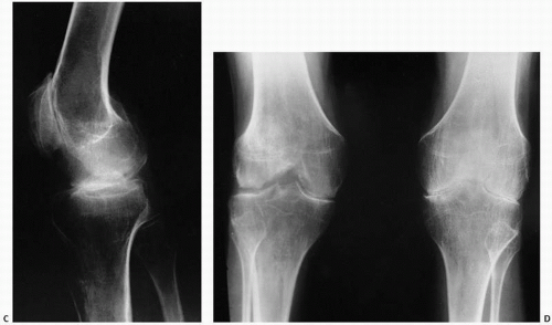
FIGURE 12.8. (Continued) (C, D) Degenerative changes of the knee.

Stage II is characterized by osteopenia in the subchondral surfaces of the affected joint. Overgrowth of the epiphyses is usually not noticeable. Joint contractures, such as fixed flexion of the knee, result in apparent enlargement of the ends of the bone on anteroposterior radiographs because of the increased distance between the knee and the x-ray cassette. This false enlargement is excluded if the lateral view is also evaluated. Enlargement of the ends of the bone is regarded to be secondary to hypervascularity, present in some cases of hemophilia. The deposition of hemosiderin in the capsule of a joint renders it slightly more dense than the surrounding soft tissues. This is most easily detected in the elbow and ankle, but it can also be noticed in the knee.

Changes in bone contour are noted in stage III. There is squaring of the patella, widening of the intercondylar notch, and subchondral cyst formation. Care should be exercised not to confuse a tunnel view of the knee (an anteroposterior view of the knee with flexion contracture is a tunnel view) with an intercondylar notch that is really widened. The subchondral cysts tend to be larger in hemophilia than in other inflammatory arthritides.

Erosion and destructive changes in the articular cartilage result in narrowing of the joint space. The changes in the articular cartilage are secondary to intra-articular liberation of digestive enzymes, as well as to prevention of the normal nourishment by synovial fluid, which cannot diffuse into cartilage coated by blood. These abnormalities are seen in stage IV.

The final stage, stage V, is defined by extensive bone destruction with marked deformity and rigidity of the joint.

The extra-articular manifestations of musculoskeletal hemophilia include muscle hemorrhage and pseudotumors. Hemorrhage in muscles can lead to neurovascular compromise. Such hemorrhage is most common in the gastrocnemius muscle and in the volar muscles of the forearm, where it can lead to a compartment-type syndrome. Bleeding in the iliacus muscle can result in femoral nerve palsy. MRI and CT are helpful in the diagnosis of episodes of bleeding into muscles.

Pseudotumors are the result of bleeding inside a bone, underneath the periosteum or in the soft tissues. This is a rare complication in which the hemorrhagic blood fails to be resorbed, accumulates, and slowly erodes the adjacent bones. It is usually an extremely painful condition and occurs in fewer than 1 percent of patients. Pseudotumors in adults are most common in the thigh, pelvis, and retroperitoneum, whereas in children they can occur in the hands and feet.

Surgical joint reconstruction in hemophiliacs has historically been necessary to preserve function. Although the mechanical survival of total knee replacement is quite good, the prevalence of infection as a complication is high (23).

Intra-articular, intrabursal, and soft tissue bleeding in hemophilia may result in painless masses clinically that roentgenographically mimic a tumor. These masses consist of spongy coagula of partially clotted blood encapsulated by thick, fibrous membranes. Complications of these so-called hemophilic pseudotumors include muscle and bone damage, infection, and neuropathies. Surgical removal is not without danger (24,25). They occur in 1 to 2 percent of hemophiliacs (26), mostly in the lower extremity and pelvis. Bleeding in the vicinity of the periosteum has been implicated in the peculiar juxtaosseous changes, which have included cyst-like bone changes and large soft tissue masses eroding bone.
MRI studies indicate a heterogeneous low-intensity signal on T1-weighted images with a surrounding capsule and septa of low signal intensity and a high-intensity signal on T2-weighted images (27).

Before 1970, hemophilia A was still associated with significant severe disability and even death at a young age. However, the median life expectancy has increased significantly throughout the 20th century from 11.4 years to 68 years in carefully followed populations (28). Nonetheless, the transfusion of blood products, including factor VIII concentrates, has led to one of the well-recognized modern complications of hemophilia—transfusion-related acquired immunodeficiency syndrome (AIDS). Life expectancy has now reversed after considerable gains. Serologically detectable antibodies to human immunodeficiency virus (HIV) developed in large numbers of hemophiliacs beginning around 1979. Approximately two-thirds of HIV-positive hemophiliacs have eventually died of AIDS.

Therapy more recently has been with factor VIII concentrates exposed to vigorous virus-killing heat treatment or solvent cleaning. Genetically engineered products are now available and are being tested for complications. Expense is a significant consideration (22).


FIGURE 12.9. Roentgenographic (A), gross (B), and histologic (C) appearance of myelofibrosis. Dense bone results from increased bone remodeling associated with paratrabecular fibrosis. The causal relationship is unknown.

In treating chronic hemophilic arthropathy, standard measures include factor replacement, physiotherapy, and operative synovectomy by conventional arthrotomy or arthroscopy. Nonoperative techniques, including the destruction of synovial tissue by intra-articular injection of radioactive agents such as colloidal 32P chromic phosphate, have been used (29).


Myelofibrosis/Myelosclerosis/Myeloproliferative Disease

Myeloproliferative disorders comprise several clonal hematologic diseases that are thought to arise from abnormal transformations in the hematopoietic stem cells resulting in the overproduction of functional or dysfunctional blood cells (30). These disorders are characterized by a long clinical course, and they may result in end-stage changes of the marrow termed myelofibrosis. The term myelofibrosis reflects the microscopically evident obliteration of red marrow by fibrosis and thickened trabecular and cortical bone (Fig. 12.9). The encroachment on marrow space results
in radiographically evident sclerosis (density) that in its terminal stages causes an inability to extract marrow for diagnosis (“dry tap”). Roentgenographic changes are seen in approximately half the cases and particularly affect sites of adult hematopoiesis (Fig. 12.10).


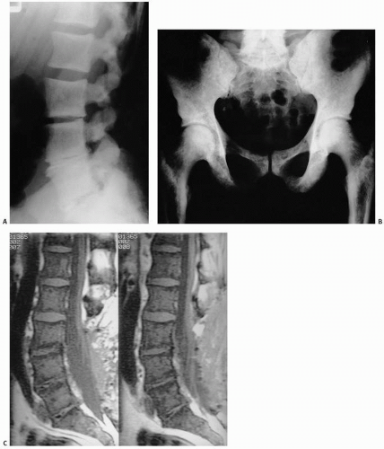
FIGURE 12.10. Radiographs of the spine (A) and pelvis and femora (B) in a patient with myelofibrosis reveal dense (sclerotic) bone. Normal bone contours are essentially maintained in the sagittal MRI. (C) The signal is low on a T1-weighted image because of replacement of the normal marrow fat by fibrosis.

The roentgenographic changes include diffuse sclerosis, sclerotic areas mixed with patchy radiolucencies, and occasionally periosteal reactions.

The contour of the bone is usually well maintained, a finding that is helpful in separating myelofibrosis from Paget’s disease. The
axial skeleton and large bones are most often affected. Malignancies that result in osteoblastic metastases, such as those of the prostate or the breast, can at times be confused with myelosclerosis.


Small Cell Malignant Tumors (Round Cell Tumors)

Tumors from which biopsy specimens reveal small cells with uniform round blue nuclei and scant cytoplasm have been referred to as small cell, round cell, or blue cell tumors (31) (Table 12.5). “Round” refers to the uniform shape of the nuclei, “blue” to the color of the nuclei on routine hematoxylin and eosin staining, and “small” to the relative lack of cytoplasm (Fig. 12.11). In fact, the “small cells” of small cell tumors are larger than most inflammatory cells.

These tumors comprise a wide range of neoplasms varying considerably in regard to prognosis, etiology, site of origin, and involvement of the skeletal system. For the most part, these are childhood cancers (Fig. 12.12).

Cancer is the leading cause of death from disease in children 1 to 15 years old (32). Estimated incidences have been reported (Fig. 12.13). However, significant improvement in the life expectancy of children with many of these tumors has been achieved (Fig. 12.13).

To achieve an accurate diagnosis, the pathologist requires adequate viable tissue on which a battery of special studies can be performed, which may include histochemical stains, immunohistochemical stains, tissue marker studies, molecular genetics studies, and electron microscopy (33) (see Appendices III and VI).


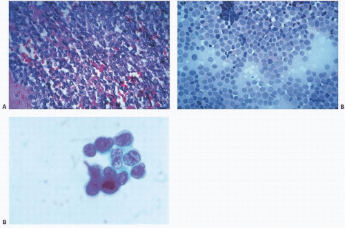
FIGURE 12.11. Small round (blue) cell tumors. “Small” refers to the size of the cells in relation to other cells and to the scant amount of cytoplasm; “blue” to the uptake of hematoxylin stain by the nucleus; “round” to its contour resulting from the nucleus being usually round. (A) Tissue section. (B) Touch imprint. (C) Cytology preparation. Increasingly finer detail is noted with each technique.


TABLE 12.5 Small Round Cell Tumors of Bone


Ewing sarcoma family of tumors


Neuroblastoma


Embryonal rhabdomyosarcoma


Desmoplastic small round cell tumor


Small cell osteosarcoma


Mesenchymal chondrosarcoma


Although the traditional morphological evaluation of bone tumors by standard light microscopy correlated with imaging findings remains the cornerstone of bone tumor diagnosis, the considerable progress that has been made in molecular pathology has enhanced our understanding of the genetic background of cancer and has led to more definitive diagnostic classification (34,35).


Ewing’s Sarcoma Primitive Neuroectodermal Tumor

Ewing sarcoma is the second most common primary malignant bone tumor occurring in children and young adults. Osteosarcoma is the most common. Ewing accounts for approximately 10 percent of all primary bone tumors. An annual incidence of 2.93 cases/1,000,000 was reported between 1973 and 2004, and the incidence has remained steady (36). Peaking in the second decade of life, it has a preference for the diaphysis and metadiaphyseal regions of long bones and may present with a painful mass. Soft tissue extension is common, but pathologic fracture much less so. Clinically,
constitutional symptoms such as fever and anemia, weight loss, and leukocytosis are common. Symptoms such as fever, laboratory findings of elevated sedimentation rate, and leukocytosis may lead to a misdiagnosis of osteomyelitis and a potentially catastrophic delay in diagnosis and treatment. A sharp and defined margin best visualized on T1 MRI images in comparison with short tau inversion recovery images has been suggested to be the most significant feature of Ewing sarcoma in differentiating it from osteomyelitis (37).


FIGURE 12.12. Small round cell tumors. (A) Neuroblastoma (note rosettes). (B) Ewing sarcoma. (C) Well-differentiated lymphocytic (mimics normal lymphocytes) lymphoma. (D) Large cell lymphoma. (E) Embryonal rhabdomyosarcoma (note wisps of red “muscle” cytoplasm).

Preferentially affecting the large long bones (in order of frequency the femur, tibia, humerus, and fibula) and flat bones (pelvis, rib, and scapula), two-thirds of cases occur in the lower extremities, pelvis, and sacrum (Fig. 12.14). Kissane et al. (38), in reviewing the Intergroup Ewing’s Sarcoma Study cases, noted the overwhelming predilection of Ewing sarcoma for the lower segment of the skeleton, with two-thirds of cases arising in a location inferior to the thoracolumbar junction. The ratios of skeletal differences are dramatic:


  • Foot to hand, 6:1


  • Leg to forearm, 69:6


  • Femur to humerus, 63:32


  • Pelvic girdle to shoulder girdle, 52:5

It is a tumor of childhood, with most cases diagnosed before the age of 20 years and the majority between 5 and 15 years of age. Delays in initial diagnosis may be due to erroneous traumatic etiologies.


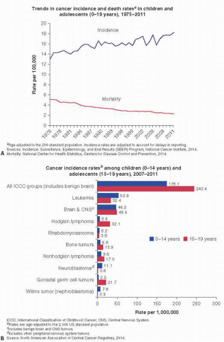
FIGURE 12.13. Cancer Facts (American Cancer Society 2015).

Showing a predilection for persons of European descent, this tumor is rare in African Americans, especially African American men. Previously considered universally fatal, Ewing sarcoma is currently curable in about half of patients who undergo combined surgery, radiotherapy, and chemotherapy.

At diagnosis, 63.8 percent of cases showed local/regional involvement, 27.4 percent distant metastases, and 8.8 percent were unstaged in the 1973-2004 SEER data (36). Fewer than 5 percent of cases involve more than one bone, and although distant metastases are noted in about 20 percent, occult micrometastases are believed to be present at the time of diagnosis in as many as 80 percent of cases (39). Ewing sarcoma is the most common source of brain metastases in children with solid tumors. Skip metastases in bone are seen in fewer than 5 percent of cases (40).

Roentgenographically, classic Ewing sarcoma presents as a permeative, poorly demarcated diaphyseal lesion with a lytic or mixed lytic-sclerotic picture (Fig. 12.15). Characteristic periosteal new bone is often layered and may resemble onion skin. Both CT and

MRI and PET scanning are better to define the extent of the tumor, with MRI particularly useful in defining marrow extension. MRI may be helpful in monitoring response to chemotherapy. Unusual roentgenographic presentations include cyst-like lesions (41) and those resembling fibrous dysplasia (42).


FIGURE 12.14. Skeletal distribution of Ewing sarcoma.


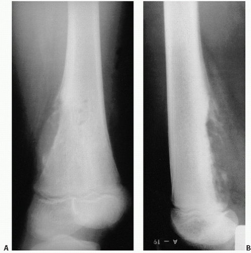
FIGURE 12.15. (A, B) Roentgenographic features of Ewing sarcoma. A lytic lesion is poorly defined, with obvious cortical destruction, soft tissue mass, and periosteal formation of new bone. The tumor is located in the junction of the metaphyses and diaphyses, and apparently spares the growth plate and epiphyses. In most cases, Ewing sarcoma occurs in the diaphyses of long bones and in flat bones. Histology of Ewing sarcoma. (Continued)


FIGURE 12.15. (Continued) (C) Sheets of round cells with bland nuclear detail and sparse, indistinct cytoplasm. (D) periodic acid-Schiff stain for glycogen shows abundant pink glycogen in the cytoplasm. (E) Touch preparation. (F) Cytologic detail of fine stippled nuclear chromatin.

Grossly, Ewing sarcoma ranges from grayish to silvery white, and is often moist and liquid in consistency, sometimes resembling an abscess. Hemorrhage and necrosis are common.

Microscopically, Ewing sarcoma consists of sheets of uniform cells with uniform round or oval nuclei, indistinct cytoplasm, and small, inconspicuous nucleoli (Fig. 12.14). Chromatin is finely dispersed. There is little, if any, intercellular stroma. Foci of necrosis are common. Mitoses may be seen, but are not frequent. A lobular growth pattern may be present, and there is often prominent capillary vasculature. Individually degenerating cells may be seen (43). On PAS staining for glycogen (which is diastase digestible), Ewing cells demonstrate abundant pink globular perinuclear positivity, a feature that facilitates the distinction from other small cell tumors of bone such as lymphoma, neuroblastoma, embryonal rhabdomyosarcoma (Fig. 12.14D). Cells, despite often being referred to as small, are large—approximately the size of histiocytes.

Tumors that show primitive neural-like architecture, with pseudorosettes and occasional spindling, have in the past been assigned to separate groups of primitive neuroectodermal tumor (PNET), but it is clear that Ewing and PNET share genetic and phenotypic features, and the differences reflect a spectrum of differentiation of the same tumor family.

Recently, unusual histologic variants include a Ewing sarcoma with epithelial differentiation in the humerus and those with features of an adamantinoma, but these lesions can be differentiated on the cytogenetic profile of Ewing, the classic translocation in Ewing t(11;22) not found in adamantinoma. In addition, adamantinoma expresses cytokeratins 14 and 19, and Ewing 8 and 18 (44).

So-called adamantinoma-like Ewing has been shown to harbor the t(11,22) (45).

The Ewing sarcoma family of tumors, which includes peripheral neuroectodermal tumor, is defined genetically by specific chromosomal translocations. These translocations include t(11;22) and q(24;q 12) (Fig. 12.16), and result in fusion of the EWS gene with a member of the ETS family of transcription factors, either FLI1, the Friend leukemia virus integration 1, (90 to 95 percent) or ERG (5 to 10 percent) (46). The resultant fusion products function as oncogenic aberrant transcription factors.

The EWS gene is a member of the TAT family of RNA-binding proteins that can activate transcription factors. The gene is expressed in all tissue types. The genes involved in the Ewing family translocation (FLI-1, ERG, etc) are in the ETS family of transcription factors that recognize a conserved DNA motif (GGAA/T) (31). The resultant chimeric protein brings the ETS family gene under the activation of the EWS component.


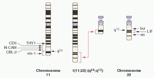
FIGURE 12.16. Translocation t(11;22) in Ewing sarcoma.

Research into the pathogenesis of the Ewing family of tumors has been hampered by lack of knowledge of the cell of origin (47), and by differences in findings using mouse cell models versus human cell models for evaluating the effects of expression of the fusion transcripts. It has been suggested that the EWS-FLI1 fusion protein may inhibit normal differentiation of the presumed pluripotent cell of origin (possibly mesenchymal stem cell), which is the precursor of chondrocytes, osteoblasts, and adipocytes. Introduction of the chimeric translocation protein into mouse marrow mesenchymal stem cells induced small round cell tumors in immunocompromised mice. Another line of evidence is that inhibition of the chimeric protein in tumor cell lines induces reversion to a mesenchymal phenotype, either adipocytic or osteogenic (48). RNAi inhibition of the fusion transcript in tumor cell lines has shown differentiation into mesenchymal cell types. It has not yet been possible to demonstrate that the translocation protein by itself is sufficient to transform human cell lines; it may be that other genetic alterations may be needed to make the cells susceptible, including aberration in the p53, INK4A, IGF-1/IGF-R, CD99, and other pathways. The function of MicroRNAs in modulating gene expression in this family of tumors has been a productive line of research (49).

EWS-FLI1 functions both as a transcriptional activator and as a repressor. Among the genes upregulated is NKX2.2, a transcription factor in the sonic hedgehog (shh) pathway. In this case, EWS-FLI1 indirectly downregulates gene expression by upregulating repressors.

The insulin-like growth factor pathway is known to be involved in supporting tumor growth in autocrine fashion (50). EWS-FLI1 inhibits the expression of IGFBP3 (IGF-binding protein 3), perhaps enhancing EGF activity. Ewing sarcoma cells express both IGF and IGF-1R (insulin-like growth factor 1 and its receptor). Clinical trials of agents interfering with this autocrine pathway are underway.

CD99/Mic2, which is an important epitope in the immunohistochemical approach to the diagnosis of the Ewing family of tumors, also represents a potential target for treatment. In vitro studies have shown that anti-CD99 antibodies induce cell death (apoptosis) in Ewing sarcoma cell lines.

Other cell cycle regulatory pathways common to many tumor types are also involved in Ewing including the retinoblastoma (RB) and p53 pathways. Cyclin D1 (a negative regulator of RB) is upregulated, and the RB protein as well as p161NK4A is often inhibited or mutated; p53 or p151NI4a/ARF mutations also carry an adverse prognostic significance.

Although these translocations have been found in some other tumors, they are considered characteristic of Ewing and represent the most important confirmatory diagnostic tool in conjunction with the histopathology as assessed by light microscopy.

Some studies have shown that different types of EWS-FLI1 fusions have prognostic significance. Patients with fusions called type 1 (EWS exons 1-7 link with FLI1 exons 6-9) may fare better (51).

These translocations are reliably identified in fixed tissue by FISH and by RT-PCR (33).

In the workup of Ewing sarcoma, it behooves the pathologist to process tissue adequately and appropriately. This includes:


  • Confirmation at frozen section or biopsy that adequate viable tumor has been sampled


  • Preparation of cytologic smears or touch imprints


  • If assessing fluid material (e.g., pleural effusions or tissue from liquefied tumor), the fluid should be centrifuged and the resultant pellet fixed with formalin prior to making a paraffin block. This material can be used for microscopic examination, immunohistochemistry, and polymerase chain reaction (PCR) and fluorescence in situ hybridization (FISH) analyses for cytogenetics.


  • A minimum of 100 mg of viable tumor snap-frozen for molecular studies


  • Formalin-fixed tissue for microscopy and immunohistochemistry


  • Sample fixed in glutaraldehyde for potential ultrastructural studies to identify cross-striations (to rule out rhabdomyosarcoma) or desmosomes (to identify epithelial features)

Immunohistochemically, Ewing sarcoma has been studied intensely (33,52,53).

The most commonly used immunohistochemical marker for Ewing sarcoma is the p30/32 MIC2 antigen, a 30- to 32-kd cell surface glycoprotein encoded by the pseudoautosomal MIC2 gene and recently associated with the CD99 antigen (54). It is recognized by different monoclonal antibodies, including HBA-71, 12E7, and 013, which has led to a plethora of confusing references (Table 12.6). Although CD99 is currently considered useful in specifically classifying Ewing sarcoma (52,53), Scotlandi et al. (54) have
shown it to be present in other cultured cell lines, including 50 percent of mesenchymal chondrosarcoma, and in some cases of small cell osteosarcoma (52), 90 percent of lymphoblastic lymphomas, 20 to 25 percent of embryonal rhabdomyosarcomas, and 75 percent of poorly differentiated synovial sarcomas. The FLI-1 protein is expressed in 84 percent of Ewing family tumors (33), but may also be seen in lymphoblastic leukemia/lymphoma and other lymphomas. Expression of neural markers such as CD57, S100, and chromogranin may be associated with differentiation toward the PNET end of the Ewing family spectrum. Other markers may be expressed such as keratin (up to 25 percent), vimentin, and desmin, emphasizing the importance of selecting the appropriate antibody panel to cover all entities in the differential diagnosis of these tumors.


TABLE 12.6 Immunohistochemical Profile of Selected Small Cell Tumors


Tumor


CD99


FLI-1


LCA


B Cell


T Cell


TdT


CK


Chr


S100


Desmin


Myogenin/MYOD1


WT1


EFT


+


+


+/-


+/-


-/+


LBL/ALL


+/-


+


+/-


+


+/-


+


+/-


NHL-other


+/-


+


+


+


+


Mesenchymal chondrosarcoma


+/-


+/-


+


+/-


+/-


Small cell OS


+/-


?


+/-


+/-


+/-


?


?


Rhabdomyosarcoma


+/-


+/-


+


+


+/-


DSCRT


-/+


+/-


+


+/-


+/-


+


+


Neuroblastoma


+/-


+


+/-


LCA, leukocyte common antigen; Tdt, terminal deoxynucleotidyltransferase; CK, pan-cytokeratin; Chr, chromogranin; EFT, Ewing family tumors; LBL/ALL, lymphoblastic lymphoma/acute lymphoblastic leukemia; NHL, non-Hodgkin lymphoma; OS, osteosarcoma; DSCRT, desmoplastic small round cell tumor; ?, not known; +, positive; -, negative; +/-, variable; -/+, rare positive.


Modified from Hameed M. Small round cell tumors of bone. Arch Pathol Lab Med. 2007;131:192-204; Gao A, Kahn LB. The application of immunohistochemistry in the diagnosis of bone tumors and tumor-like lesions. Skeletal Radiol. 2005;34:755-770.


Currently, demonstration of EWS/FLI-1 or variant translocations is the gold standard for the diagnosis of Ewing family tumors, with immunohistochemistry as a vital adjunct.

Confirmation of the diagnosis by molecular techniques is usually done by either FISH or RT-PCR. FISH has the advantage of permitting identification of the EWS rearrangement irrespective of the fusion partner. RT-PCR, which requires specific primers for each gene partner, allows the identification of the exact fusion partner.

As with all chemotherapy-treated and subsequently resected tumors, the pathologist plays an important role in assessing the degree of tumor necrosis. In general, it is of clinical usefulness if an assessment is made of the percentage of the tumor that is necrosed:


  • 0 percent necrosis: No chemotherapy effect = Grade I


  • <50 percent necrosis: Partial or low chemotherapy effect = Grade IIA


  • 50 percent to 95 percent necrosis: Partial or high chemotherapy effect = Grade IIB


  • 96 percent to 99 percent necrosis: Only scattered viable foci of tumor = Grade III


  • 100 percent necrosis: No residual viable tumor after extensive sampling = Grade IV

The prognostic importance of assessing necrosis has been proposed with 3-year survival percent at 30 percent if there is no or less than 10 percent necrosis, 49 percent survival with 11 to 90 percent necrosis, 73 percent survival with 99 percent necrosis, and 100 percent survival if there is complete necrosis (51).

Ewing is staged on the basis of tumor size (8 cm being the cutoff), involvement of lymph nodes, and assessment of metastases. Ewing is always classified as a high-grade tumor.

Plain film radiography, CT, and MRI of the primary tumor all play a role in providing valuable staging information. Pain films can document the permeative and moth-eaten destructive pattern over time. CT can determine the integrity of the cortex and thus potential for pathologic fracture (10 to 15 percent of cases). MRI is useful in defining the extent of marrow and soft tissue spread. Metastatic disease usually to lung and bones can be evaluated with 99mTc bone scans or PET scans.

With regard to the staging and restaging of Ewing sarcoma, the sensitivity, specificity, and accuracy of both FDG PET and PET/CT are high (55). CT scans of the chest, abdomen, and pelvis are also used to assess disease at those sites.


Grading

Primitive neuroectodermal tumor/Ewing sarcoma (either intraosseous or extraosseous) is classified as high grade, and hence stages 1A and 1B are excluded for PNET/ES.

In general, favorable prognostic factors in Ewing include:


  • Age less than 17


  • Extremity involvement (distal > proximal)


  • Appendicular versus axial tumors (56)


  • Tumors < 8 cm (57)


  • Nonmetastatic tumors


  • Type I EWS-FLI1 fusion transcript


  • >90 percent necrosis following chemotherapy


  • Patients treated with surgical removal of the involved bone (39)


TABLE 12.7 Poor Prognostic Factors in Ewings Sarcoma


Fever


Anemia


Age >17 y


Tumors >8 cm or greater than 100 mL tumor volume


Metastases at time of initial presentation (20% vs. 69% event-free survival)


Pelvic and sacral tumors


Type II EWS fusion transcript


<90% necrosis after chemotherapy


>20% cells overexpressing p53


Mutations in INK4a gene


Deletion of p16 and/or INK4a


Deletion of the STAG 2 gene


Extraosseous soft tissue extension


Strand-like (filigree) histologic cellular pattern? (36)


Serum lactate dehydrogenase (? related to tumor size)


Postradiation medullary tumor


Postinduction CT findings of medullary involvement, cortical destruction, lysis, permeation, and unhealed pathologic fracture (51)


Time of first recurrence


Concomitant recurrence at local and distant sites


Other debatable poor prognostic factors are high levels of lactate dehydrogenase, tumor volume >100 mL, and >20 percent of cells expressing p53 (Table 12.7).

With current therapy, the survival rate for localized disease is approximately 70 percent (58). However, the survival for recurrent or metastatic disease remains poor.

18F-FDG-PET scanning has been widely used to measure response to chemotherapy. Whereas many studies consider a reduction of the SUV max (maximum standard uptake value) to below a value of 2 or 2.5 as the appropriate cutoff value to define good responders, other measurements have been used. For example, a 90 percent reduction in the metabolic tumor volume (MTV as defined as the number of voxels within the volume of interest [VOI] that had an uptake greater than that of the chosen background threshold) has been suggested as a good response (59).

In recent decades, the prognosis of localized Ewing sarcoma has improved dramatically, a fact widely accepted to have been brought about by systemic chemotherapy. In the first Intergroup Ewing’s Sarcoma Study (IESS-I), from 1973 through 1978, four-agent chemotherapy led to a 5-year relapse-free survival in 56 percent of cases (31). However, children with pelvic tumors did worse (less than 25 percent 5-year survival). In IESS-II, through 1982, 5-year survival reached 73 percent. Relapses included local disease in 9 percent and metastases to the lungs and bones. Some studies indicate better survival in patients younger than 10 seen initially with metastases, probably reflecting more favorable primary sites, such as the rib (60). Because 50 percent of local failures occur centrally within fields of radiation rather than peripherally, there is renewed interest in including surgical resection when feasible.

Only gold members can continue reading. Log In or Register to continue

Stay updated, free articles. Join our Telegram channel

Jul 24, 2016 | Posted by in PATHOLOGY & LABORATORY MEDICINE | Comments Off on Bone Manifestations of Hematologic Disorders and Small Cell Tumors

Full access? Get Clinical Tree

Get Clinical Tree app for offline access