Bone-Forming Bone Tumors and Tumor-like Lesions


Bone-Forming Bone Tumors and Tumor-like Lesions


Osteoma

Osteomas are benign slow-growing radiodense tumor-like lesions characterized pathologically by predominantly mature lamellar bone. Several clinicopathologic syndromes are known (Table 9.1).

Osteomas may particularly affect the skull and facial bones, especially the mandible and frontal and ethmoid sinuses. Dome-shaped ivory-like excrescences on the inner or outer surface of the calvarium or protruding into the orbit or paranasal sinus are typical (Fig. 9.1). Clinically, a painless mass is noted. Frontal, ethmoid, maxillary, and sphenoid sinuses are the ones most frequently affected, and in that order (1). Sinusitis, nasal discharge, headache, pain, or loss of smell may be the presenting symptom. In the frontoethmoid sinuses, large growths may erode the dura and cause cerebrospinal fluid leakage, pneumatocele, meningitis, or cerebral abscess. Orbital lesions may produce exophthalmus, double vision, and even loss of vision.

Earwaker (2) has estimated the incidence of paranasal sinus osteomas as 3 percent, with a predilection for the fifth and sixth decades. The frontal sinus was commonly involved. Most osteomas are usually less than 5 cm.

The prevalence of osteomas in the skull has been studied, showing that 5 percent of patients in a consecutive autopsy series contained meningeal osteomas located at the dura-falx junction. These lesions were seen in higher association with renal disease and consisted of more extensive remodeling than usually encountered in the classic compact osteoma (3). Patients had abundant osteoid and numerous osteoclasts, such active bone remodeling known to be associated with renal disease. It is most likely that some nodular densities reported in the radiology literature as “calcification” in renal disease represent a type of incipient osteoma.


TABLE 9.1 Osteoma: Clinical and Roentgenographic Types


Enostosis (osteoma-like growth within bone)a


Osteomas involving the skull and facial bones


Long bone osteomas


Parosteal osteomas


Gardner syndrome


Osteomas associated with systemic illness


a See Chapter 2, Figures 2.32 and 2.33.


Parosteal osteomas (or long bone osteomas) are benign radiodense growths on the surface of bone composed of essentially cortical-type haversian bone (4). Its importance rests in the distinction from parosteal osteosarcoma, which is characterized by the coexistence of bone and a cellular fibrous tissue. Parosteal osteomas are usually reported in the lower extremity (4) (Fig. 9.2). Patients are usually in the fourth or fifth decade.

Roentgenographically, a dense or oval-shaped mass is noted contiguous with the cortical surface of the bone. There is no periosteal reaction, and there are no areas of radiolucency. The surface is smooth or smoothly lobulated. The underlying cortex of the host bone may be slightly thickened, and cross-sectional studies may show slight encroachment of medullary bone, but these changes are minimal if present.

Because of the difficulty in distinguishing this lesion from osteosarcoma, some recommend definitive surgical removal even when the diagnosis of osteoma is likely (5).

Osteomas of the mandible, calvarium, and even long bones are seen in association with colonic intestinal polyps in Gardner syndrome, which is also characterized by odontomata, supernumerary, and unerupted teeth, and soft tissue tumors, including fibromas, retroperitoneal and mesenteric fibromatosis, and epidermal inclusion cysts. Gardner syndrome is an autosomal dominant genetic disorder and is of particular importance because of the associated malignant change seen in the adenomatous lesions of the intestine. First described in 1953 by Gardner and Richards (6), the classic entity is considered to arise from each of the three germinal layers in contrast to familial polyposis coli, an endodermal defect. Classically, a clinical triad is noted: (a) gastrointestinal polyposis,
(b) osteomas, and (c) epidermal inclusion cysts of the skin. Osteomas in Gardner syndrome may or may not be well defined and has been reported in long bones, skull, mandible, and paranasal sinuses. Defective dentition and fibrous tumors may also be present. Polyposis in Gardner syndrome begins late in childhood or early in adulthood, and often results in gastrointestinal carcinoma.


FIGURE 9.1. Exophthalmos of the right orbit (A) because of a large, well-circumscribed, ivory-white osteoma of the frontal sinus (B).


FIGURE 9.2. Skeletal distribution of parosteal osteoma. (From Bertoni F, Unni KK, Beabout JW, et al. Parosteal osteoma of bones other than of the skull and face. Cancer. 1995;75:2466-2473. Copyright © 1995, American Cancer Society. Reprinted with permission of Wiley-Liss, Inc.)

Grossly, osteomas are dome shaped or lobulated, dense and hard, with the appearance and texture varying with the degree of active growth (Fig. 9.3). Four types have been described and have been summarized by Earwaker (2): (a) ivory (eburnated)—hard, dense mature bone with no definable haversian systems; (b) compact—compact lamellar structure with haversian canals;

(c) spongiosis—periphery of compact bone with radial septa and intervening marrow spaces; and (d) mixed—bone and fibrous tissue. The characterization into these subtypes is not clinically relevant.


FIGURE 9.3. Osteoma. Gross and microscopic. An ivory-white osteoma appears nipple-like over the smooth calvarium of the skull (A); histologically, the lesion appears as a mature bone blending imperceptibly into the adjacent outer table of the skull (B); osteomas are usually mature cortical bone (C, left), but, while forming, will show foci of remodeling (C, right—open space). Osteoblastoma-like osteoma. Irregular spicules of bone in a fibrovascular stroma with active osteoblast and osteoclast remodeling. [(D) Roentgenograph; (E1 and E2) Microscopy].

Osteomas are often covered by a thin glistening membrane. Osteomas usually consist of circumscribed foci of dense bone contiguous with host bone. They consist primarily of mature lamellar cortical-type haversian bone. Wide zones of either cortical or trabecular bone may be seen, and, although usually mature lamellar bone predominates, immature woven bone may be noted. There are sparse interosseous spaces, which may be filled with vessels, fibrous tissue, fat, or even bone marrow. In general, the typical osteoma has a lobulated appearance on microscopy, and consists of mature remodeling haversian bone and abuts rather than blends into the surrounding tissue. Active remodeling of bone in an osteoma showing numerous osteoblasts and osteoclasts may mimic an osteoblastoma, the so-called osteoblastoma-like osteoma (Fig. 9.3 D,E).

In general, osteoblastomas form expansile, partially calcified tumors, whereas osteoblastoma-like osteomas are heavily ossified masses that microscopically have much more mature bone in the form of solid, compact bony tissue. They do not appear to act more aggressively than osteomas without the osteoblastoma-like features (1).

The etiology of osteoma is obscure. The lesion does not recur if surgically excised, and is not associated with malignant change. Theories of etiology include hamartomatous, inflammatory episodes eventuating into osseous repair, or even a sclerotized end-stage of fibrous dysplasia favored by Jaffe.


FIGURE 9.4. Skeletal distribution of osteoid osteoma.


Osteoid Osteoma

Osteoid osteoma is a small solitary, benign bone-forming lesion within host bone that has characteristic clinical, roentgenographic, and pathologic features (7,8). Pain, often severe at night, and relieved by aspirin, with a hot, well-circumscribed lesion on bone scan, is characteristic. Symptoms vary from a mild ache to severe debilitating pain. Osteoid osteomas characteristically occur as a well-defined nidus of remodeling spicules of cancellous-type bone within the cortex of the host bone. Less commonly, it is seen in the cancellous marrow bone and even less frequently in a subarticular or juxta-articular location.

Clinically, osteoid osteoma has a peak occurrence in the pediatric population age 11 to 20 years, with 90 percent of cases occurring between ages 5 and 30 years (Fig. 9.4). There is a male predominance of 2:1.

The presenting symptom, characteristically, is pain. However, clinical manifestations that are sequelae of the particular host bone involvement include, in the extremities, muscle wasting and limping; in the spine, scoliosis (9), disc problems, and neurologic symptoms; and at peri-epiphyseal sites, arthritis and skeletal asymmetry. Osteoid osteoma is the most common cause of painful scoliosis in an adolescent.

Juxta-articular osteoid osteomas may be accompanied by negative plain radiographs, but associated synovitis may mimic an inflammatory arthropathy such as rheumatoid arthritis (10).

The explanation of pain in osteoid osteoma has received some attention. Unmyelinated nerve fibers have been demonstrated
within the nidus (11). The relief of pain by aspirin is thought to be due to the prostaglandin-inhibiting action of aspirin, prostaglandins having been linked to lesional tissue in osteoid osteoma (12). Prostaglandins E2, F, and alpha have been reported in much larger-than-expected quantities by Makley and Dunn (12). Their link to pain may be either through vasodilatory effects with local blood vessel proliferation causing a pressure-type effect or through an effect on the bradykinin system (13). Prostaglandins may in fact produce or contribute to the production of osteoid osteoma, prostaglandin receptors having been found in bone cells. The remodeling bone effects of prostaglandins are well known and witnessed in other conditions such as mastocytosis (see Chapter 4).

Although osteoid osteoma has been reported in most bones, there is a predilection for the lower extremities, with half the cases involving the femur and tibia.

Osteoid osteoma has a classic roentgenographic appearance (Fig. 9.5). Although only poorly circumscribed cortical bone sclerosis may be noted, a centralized radiolucent nidus may be detected within the sclerotic cortical bone. Sclerosis may obscure the nidus on routine radiographs. Computed tomography (CT) scanning may detect the nidus, whereas routine radiographs only show sclerosis. SPECT-CT is used by some to amplify the presence of the nidus and improve diagnosis. The lesion is usually less than 1 cm in size. Hypervascularity and abundant mineralization in osteoid osteoma explains the typical hot bone scan.

Bone scans may demonstrate the “double density” sign. This refers to the central occurrence within a hot scan focus of an even hotter focus. The hotter focus is the actual osteoid osteoma nidus. The less hot focus is the surrounding reactive tissue.


FIGURE 9.5. Osteoid osteoma. Roentgenographic appearance. (A) The left distal tibia shows distinct cortical sclerosis. (B) Tomographic films demonstrate a radiolucent nidus. (C) The lesion on CT scan shows cortical sclerosis and central nidus, within which is a radiodense (bone-forming) central core. (Continued)

Periosteal reactions are common. Osteoid osteomas located in intra-articular sites do not elicit a reactive sclerosis and may be obscure (Fig. 9.5E).

Although magnetic resonance imaging (MRI) has been used in detecting osteoid osteoma, relying on it can be misleading (14). Reactive bone changes, soft tissue changes, and bone marrow edema can all interfere with proper diagnosis.

Grossly, osteoid osteoma may be difficult to identify because they are often small bone foci within sclerotic bone. Large lesions are reddish in color (Fig. 9.6). The zonal architecture is characteristic (Fig. 9.7).

The lesion is usually intracortical, where a well-circumscribed round or oval nidus consists of interlacing spicules of cellular remodeling cancellous (trabecular) bone (Fig. 9.8). The bone spicules are replete with bone lining cells including osteoclasts and numerous osteoblasts. Osteoid surfacing the bone is abundant. Between spicules is a richly vascular loose fibrovascular tissue. Cartilage, bone marrow elements, and mitoses are usually not present. The nidus is characterized by more abundant mineralization centrally (a central sclerosis on x-ray) and separation from host bone peripherally by a fibrovascular, sparsely mineralization perimeter.

Although the etiology of osteoid osteomas remains obscure, it causes pain and may require surgical excision. In some cases of clinically suspected osteoid osteoma, the nidus is not found (15,16) (Fig. 9.9). Failure to surgically localize the lesion or identify it pathologically in multiple submitted specimens is a probable explanation (Fig. 9.10). Because bone scanning with 99mTc-labeled diphosphonate compounds has been used to localize a broad range of osseous lesions, including osteoid osteoma (17), a preoperative
technetium injection can localize the lesion intraoperatively. Coupled with intraoperative localization by scintillation probe, conservative surgical excision can be accomplished and is of considerable benefit to the patient (Table 9.2).


FIGURE 9.5. (Continued) (D) Lesions are very hot (black) on bone scan (rib). (E) CT scan. After double-contrast arthrography of the hip (the needle for the arthrogram is still in place), the nidus is seen in the anterior surface of the femoral neck. Small collection of calcium (bone) is present in the center of the lucent nidus. The lack of surrounding sclerosis is due to the intra-articular location of the lesion.

Other techniques for operative localization of osteoid osteoma include preoperative use of tetracycline (18). Tetracycline autofluoresces and, like technetium, concentrates in mineralizing tissue. Using equipment to detect the fluorescence in the operating room has been successful. In the tetracycline technique, the patient ingests 750 to 4,000 mg of tetracycline (4 mg/kg) in four equally divided doses 1 to 2 days prior to surgery. A Woods lamp is used intraoperatively to illuminate the tetracycline-drenched tissue presumed to be the nidus. Success with this technique is variable (8).

The treatment of osteoid osteoma is initially conservative with pain medication (typically nonsteroidal anti-inflammatory drugs [NSAIDS]), but surgical removal may be necessary. Curiously, as with so many pathologic lesions of the skeleton, spontaneous regression and even disappearance of the lesion has been reported.

Newer percutaneous approaches to treatment have been developed to overcome the problems associated with traditional surgery (longer anesthesia, longer inpatient time, more tissue exposure, more tissue injury, and longer recovery period).

These newer procedures include:


  • Image-guided cryotherapy (19)


  • Interstitial laser photocoagulation (20)


  • CT-guided drilling with ethanol injection


  • CT-guided radiofrequency ablation (21)


  • Magnetic resonance-guided focused ultrasound (MRgFUS) (22).


FIGURE 9.6. Osteoid osteoma (gross). Lesions consist of a well-defined focus of bone (nidus) surrounded by a reddish perimeter of less dense bone, all enveloped by dense sclerotic host bone (A, B). Large lesions have a gritty, cherry-red appearance (C).


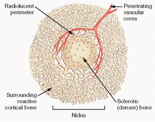
FIGURE 9.7. Osteoid osteoma. The radiolucent nidus consists of a sclerotic dense central core within a less dense perimeter of bone. The nidus is surrounded by very sclerotic host bone.


FIGURE 9.8. Osteoid osteoma (microscopy). Identified by its small size and circumscription, osteoid osteoma is often buried within thickened cortical bone (A). The nidus (B, left) is distinct from the surrounding remodeling bone (B, right). (Continued)


FIGURE 9.8. (Continued) Osteoid osteoma nidus. The nidus is characterized by thinner sinewy streams of interlacing, often incompletely mineralized, trabecular bone rimmed by abundant osteoid and osteoblasts (C, D). Osteoblasts are often irregularly shaped. Osteoclasts are also seen. Intervening stroma is sparsely cellular and strikingly vascular, often fibrovascular in nature.


FIGURE 9.9. Osteoid osteoma. Experience in a series of operations attempting to localize the lesion. (Modified after Sim FH, Dahlin DC, Beabout JW. Osteoid-osteoma: diagnostic problems. J Bone Joint Surg Am. 1975;57:154-159.)


FIGURE 9.10. Osteoid osteoma. Minute lesion measuring just a few millimeters identified by serially sectioning and x-raying the removed specimens.

Cryoablation utilizes the delivery of room temperature argon gas through a sealed insulated probe (19). Rapid expansion of the gas leads to cooling via the “Joule-Thompson” effect. With temperatures reaching over -100 °C at the tip of the probe, an ice ball is formed with the resultant crystals from the ice causing cell damage.

The advantage of MRgFUS is that it is totally noninvasive and eliminates, unlike CT-guided procedures, radiation exposure.

Best used in extraspinal locations, radiofrequency ablation works by inserting an electrode into the nidus and then heating the tip of the electrode to 90 °C for about 5 minutes. The main disadvantage is lack of histologic confirmation of the diagnosis. Skin and soft tissue burns and skin necrosis have been reported (21).

A disadvantage of radiofrequency ablation is the potential to damage nearby neurological structures, thus limiting its application in spinal locations.

Since the generation of heat in these procedures can cause tissue damage, formulae have been developed to help guide and predict temperature in the bone (23). Not surprisingly, significantly higher temperatures are found in lesions in cancellous bone versus cortical bone.


TABLE 9.2 Results of Procedures Used to Localize and Diagnose Osteoid Osteomaa


Case


Site


No. of Specimens and Designations Used


Microradiograph


Autoradiograph Image


Histopathologic Nidus


I


Finger


3; Ab, Bb, Cb


Nondiagnostic


Positive C >> A


C


II


Distal femur


2; Ab, Bb


Diagnostic


Positive A >> B


A > B


III


Femoral neck


2; Ab, Bb


Nondiagnostic


Positive B


B


IV


L4 vertebra


10; A, B, C, D, E, F, G, H, I, J


Nondiagnostic


Positive H >> I > G


H > I


V


Proximal tibia


2; A, B


Nondiagnostic


Positive A >> B


A >> B


aIn five cases, microradiographs of the specimen revealed a nidus in only one. However, after preoperative injection of technetium, localization by operative scintillation probe identified the nidus in all cases.


b Each specimen contained multiple small pieces.


Modified after Vigorita VJ, Ghelman B. Localization of osteoid osteomas—use of radionuclide scanning and autoimaging in identifying the nidus.


Am J Clin Pathol. 1983;79:223-225.


Arthroscopically assisted radiofrequency ablation has been employed for intra-articular locations such as the hip that otherwise would require a difficult approach and jeopardize damage to cartilage and bone (24).

In interstitial laser photocoagulation, bare optical fibers are inserted into the nidus and then treated with low levels of power (typically 2 to 4 W) for several minutes, creating a zone of coagulative necrosis (20).

Although usually solitary, synchronous multifocal osteoid osteomas have been described (25).


Osteoblastoma

Osteoblastoma is a benign bone-forming lesion that occupies an often tenuous niche between its clearly benign smaller counterpart, the osteoid osteoma, and its clearly malignant counterpart, osteosarcoma (Fig. 9.11). Its focal resemblance microscopically and roentgenographically to various other entities such as aneurysmal bone cyst (ABC), giant-cell tumor, osteoid osteoma, and osteosarcoma raises considerable diagnostic dilemmas (26). It is best characterized by descriptions of its prevailing salient clinical and pathologic characteristics.

The classic osteoblastoma is seen as an expansile lytic lesion in the posterior elements of the upper vertebrae of a 19-year-old man who is experiencing local pain. Pain is almost always present, as is tenderness. Pain usually increases with motion and is often radicular in nature. Scoliosis is often seen. Symptoms are thought to be related to a local mass effect. It is rare after 50 years of age.

Tissue is reddish and granular and often vascular. The microscopic appearance is usually sharply defined with clear circumscription at low power. There is a seemingly haphazard array of interlacing bone trabeculae lined by abundant osteoblasts and admixed with varying degrees of lacelike or sheeted osteoid and fibrovascular tissue (Fig. 9.12). Cartilaginous tissue without a fracture is usually not seen, but has been reported (27). Atypical mitoses are rare, but minimal mitotic activity can be seen in as many as 90 percent of cases (28).

Osteoblastoma has a predilection for the axial skeleton and spine in particular, with roughly equal distribution in cervical, thoracic, and lumbar spine (Fig. 9.13). Roughly 40 percent of cases occur in the spine, especially the posterior elements (arch and spinous processes); 30 percent affect the long bones (especially from the femur to the tibia). Epiphyseal involvement is rare. Although the epicenter of the lesion is usually within medullary bone, both cortical and periosteal osteoblastomas are well described.

Multifocal patterns of osteoblastoma have been described in as many as 14 percent of cases (28). These may involve multiple sites within the same bone (5). Kyriakos et al. (29) have coined the term “osteoblastomatosis” for a rare variant in adults composed of multiple osteolytic lesions in a unilateral distribution.

The roentgenographic appearance of osteoblastoma is quite diverse (Fig. 9.14). Predominantly radiolucent circumscribed lesions with expansile margins may be seen or lesions with more evident punctate or diffuse radiodensities. In the appendicular skeleton, 65 percent are cortical, 30 percent medullary, and 5 percent surface in location (28). Ten percent have a malignant appearance, with 25 percent showing cortical destruction. In the vertebral skeleton, involvement of the vertebral body alone is rare. Most cases involve dorsal elements. A balloon-like periosteal shell may be best appreciated on CT or MRI. The typical MRI pattern is as follows: T1 low signal and T2 high signal transversed by linear low signals (bone).

In an appendicular bone, the presence of a lytic metadiaphyseal lesion containing matrix mineralization with prominent associated surrounding edema on MRI is thought to be highly suggestive of osteoblastoma (30).

The rare cortical osteoblastomas, like their osteoid osteoma counterparts, can evoke a significant sclerosis.

Anatomic features of aneurysmal bone may be seen in as many as 16 percent of osteoblastomas (28).

The natural course of osteoblastoma is to be progressive in growth with a tendency to recur. Recurrences have been reported in as high as 21 percent of cases (28).


FIGURE 9.11. Osteoid osteoma versus osteoblastoma versus osteogenic sarcoma.

Osteoblastomas are often cured by initial therapy that consists of curettage with or without bone grafting. To avoid recurrence, a marginal resection, where feasible, is to be considered. Tumor-free margins are consistent with a prolonged disease-free interval. In surgically challenging sites such as the spine, preoperative embolization followed by “margin- free” surgery has been employed (31).

Osteoblastomas are often cured by initial therapy, which includes curettage (with or without bone grafting) for weight-bearing bones and compete resection for bones more surgically accessible. In general, en bloc resection is adequate.

In recent years, radiofrequency ablation has been found efficient by some in treating osteoblastomas including spinal osteoblastomas (32).

Osteoblastomas can present with significant systemic symptoms including fever, anorexia, and weight loss. Referred to as “toxic” osteoblastomas, they may be associated with marked periostitis and can mimic osteogenic sarcoma on imaging (33).

The differential diagnosis of osteoblastoma includes osteoid osteoma and osteogenic sarcoma. Like osteoid osteoma, local pain is common, although not, as in osteoid osteoma, typically relieved by aspirin (13,21,28) (Table 9.3). As in osteoid osteoma, symptoms such as a limp, muscle wasting, scoliosis, or neurologic symptoms may be manifestations of the affected bone. Analogies with osteoid osteoma have been made primarily on the basis of the similar, although not identical, histology and the rarely documented reports of previously diagnosed osteoid osteomas acting subsequently more aggressively.

Microscopically and roentgenographically, osteoid osteoma has a more definitive zonal architecture or “nidus” architecture as previously described.

Osteoblastoma is one-fifth as common as osteoid osteoma and usually greater than 2 cm, the usual cutoff for osteoid osteoma.

Dorfman and Weiss (34) have classified osteoblastic tumors of uncertain malignant potential into four categories: (a) low-grade osteosarcoma resembling osteoblastoma; (b) osteoblastoma transforming into osteosarcoma (35); (c) pseudosarcomatous osteoblastoma, a benign lesion in which a degenerative atypia mimics a sarcoma (36); and (d) locally “aggressive osteoblastomas,” which do not metastasize.

The latter group, aggressive osteoblastomas, is characterized clinically by local aggressive behavior and bizarre microscopic features, blurring the distinction with osteosarcoma (37). Nonetheless, these latter lesions are considered different vis-à-vis prevalence microscopically of large, plump epithelial-like (epithelioid)
osteoblasts, with deeply staining eosinophilic cytoplasm found in aggregates or sheet-like proliferation. Nuclei may be quite large, with vesicular finely clumped chromatin. Stromal mitoses are noted as well as a disorganized pattern of osteoid and osteoclasts. Cartilage and calcified cartilage are not usually encountered. In differentiating osteoblastoma from osteosarcoma, osteoblastoma stroma always has a benign fibrovascular appearance in the intervening spaces between bone production.


FIGURE 9.12. Osteoblastoma (microscopy). Interlacing spicules of osteoid and irregular and mineralized cellular bone with abundant osteoblasts (A) and intervening hypervascular stroma (B). More aggressive osteoblastomas are characterized by “epithelioid” osteoblasts, plump epithelial-like osteoblasts (C). In osteoblastoma-like osteosarcoma, lesions are less well circumscribed (D) and, on high-power microscopy, reveal more nuclear pleomorphism than in osteoblastomas (E).

The predominant cells have been shown to be similar to osteoblasts by electron microscopy (38). They contain round nuclei with well-developed rough endoplasmic reticulum and some cytoplasmic dense bones surrounded by thick matrix. The larger epithelioid cells had fewer organelles but identifiably dilated rough endoplasmic reticulum with many polysomes and ribosomes and some mitochondria.

Aggressive osteoblastoma remains somewhat controversial as an entity, some favoring that it be interpreted as an osteosarcoma (37). Nonetheless, cases reported in the literature are mostly in the spine, pelvis, and femur, but are reported throughout the skeleton unlike classic conventional osteosarcoma (Table 9.4). Mean age is 30 years, which is also different from conventional osteosarcoma (38). Osteosarcomas with radiographic and histologic resemblance to osteoblastoma can be a particularly difficult diagnostic challenge (vida infra) (30).


FIGURE 9.13. Skeletal distribution of osteoblastoma.


FIGURE 9.14. Osteoblastoma. Roentgenographic appearance. Radiodense destructive mass destroying the pedicle and transverse process of the right side of the lumbar fourth vertebra (A). Poorly defined sclerotic mass of the proximal ulna (B). Mildly expansile partially calcified lesion in the lamina of a lumbar vertebrae (mimicking a “giant osteoid osteoma”) (C).


TABLE 9.3 Clinical Differentiation: Osteoid Osteoma and Osteoblastoma (Based on 860 Osteoid Osteomas and 184 Osteoblastomas)


Osteoid Osteoma


Osteoblastoma


Age


87% ≤ age 30 y


91% ≤ age 30 y


Incidence


Relatively common


Rare


Sex ratio (M/F)


2.2:1


1.9:1


Duration until diagnosis


≤11.3 mo (average)


≤12.3 mo (average)


Pain


Moderate/marked (>98% painful) Nocturnal pain relieved by aspirin


Mild/moderate; less aspirin relief (>98% painful)


Location


Femur and tibia, 49.4%


Spine, 35.8%


Size


<1 cm


>2 cm (range, 1.0-15 cm; mean, 3.6 cm)


X-ray findings


Sclerosis, “nidus”; typical appearance


Erosive, expansile; variable appearance


Clinical course


Static (regression in approximately 12 reported cases); limited growth, if any


Enlargement; progression (no regression reported)


Treatment resultsa


4.5% recurrence


>10% recurrence (five cases sarcoma)


a After varying modes of treatment ranging from complete en bloc resection to punch biopsies only, with and without bone grafts and/or radiation therapy. Modified from Jackson RP, Reckling FW, Mantz FA. Osteoid osteoma and osteoblastoma. Similar histologic lesions with different natural histories. Clin Orthop. 1977;128:303-313 and Lucas DR, Unni KK, McLeod RA, et al. Osteoblastoma: clinicopathologic study of 306 cases. Hum Pathol. 1994;25:118-134.


Osteosarcoma


Classic (Medullary Epicenter)

Osteosarcoma is one of the most common malignant tumors of adolescence, exceeded by only leukemias, brain tumors, and lymphomas (39). There are roughly 8.2 cases per million population, with less than 1,000 new cases each year in the United States. It is a highly malignant tumor, which, by definition, produces neoplastic osteoid or bone or both. Osteoid is type I collagen, which, in the normal sequence of events, being bathed in bone-forming proteins, will form bone. Osteosarcoma characteristically arises within the metaphysis of the long bones that are the sites of the most rapid growth and greatest blood flow: the distal femur, proximal tibia, and proximal humus. It grows circumferentially through the cortex into the soft tissue raising the periosteum. It rarely invades the joint space. Fifty-six percent of all osteogenic sarcomas occur at the knee, resulting in its being the most common primary osseous knee tumor reported in the literature (32 percent) (Fig. 9.15). Of osteogenic sarcomas of the knee, 64 percent occur in the distal femur, 32 percent in the proximal tibia, 4 percent in the proximal fibula, and less than 1 percent in the patella.

Given the predilection of osteosarcoma occurring around the knee, the initial misdiagnosis of an athletic injury such as a meniscal lesion at this site has been shown in several studies (40).

Osteogenic sarcoma, for which there is an animal model in the Great Dane, has a seeming predilection for areas of rapid growth (41). It peaks, as mentioned, in the adolescent growth spurt, and is also seen with increased incidence in bone affected by Paget’s disease.

Osteogenic sarcoma characteristically occurs at the adolescent growth spurt, with the peak age of occurrence between 10 and 20 years of age; 75 percent of all cases occur between 10 and 30 years of age. There is a male predominance of 1.5:1. In the immature skeleton with an intact growth plate, the epiphysis may act as a relative barrier to its growth. Microscopic and MRI evidence of transepiphyseal spread is more common than appreciated on routine roentgenograms. Osteogenic sarcoma typically presents with pain, which is often mild and intermittent initially, but more continuous and exacerbated by deep palpation later. Pain is the most common presenting symptom occurring in about 80 percent of patients. It is usually exacerbated by activity with less than one quarter of patients showing pain at night. A mass or a swelling may be felt. Patient may present with a pathologic fracture. In general, patients are symptomatic for several weeks before coming to clinical attention. On examination, a palpable mass may be felt. Large lesions may lead to overlying venous engorgement or edema.

The laboratory workup of osteosarcoma would include alkaline phosphatase (almost always elevated) and lactate dehydrogenase (may be elevated). imaging workup of osteosarcoma includes biplanar radiographs, CT, and Prebiopsy MRI of the involved site. A whole-body three-phase bone scan should also be done.

A postbiopsy chest CT is employed to look for metastatic disease.

The characteristic radiograph reveals a radiodense or mixed radiolucent and radiodense lesion over the metaphysis with indistinct borders and periosteal elevation (Fig. 9.16). The raised periosteum creates a triangle (referred to as Codman triangle), whose borders are the intact cortex, the tumor, and the periosteum proper. CT scans may be helpful in defining soft tissue or joint penetration, with MRI defining the exact extent of involvement of the cancellous and medullary bone and soft tissue.


TABLE 9.4 Aggressive Osteoblastoma versus Osteosarcoma


Aggressive Osteoblastoma


Osteosarcoma


Age (mean), years


30


15


Site


Vertebrae


Extremities


Periosteal new bone formation


Minimal


Abundant


Nidus, i.e., circumscribed


Yes


No


Microscopic architecture


Zonal Peripheral maturation


None


Atypia


+


++


Mitoses


0/+


++


Atypical mitoses


0


+


Marrow and bone permeation


Minimal


Marked


Trabecular lining


Single cells


Often sheets


Cartilage


Minimal if at all


May be abundant


Metastases


0


+


FIGURE 9.15. Skeletal distribution of osteosarcoma.

By definition, osteosarcoma is bone forming. However, bone production may not be roentgenographically evident in all cases. Although blastic lesions predominate, mixed lesions with patchy lysis and sclerosis admixed may be seen. Telangiectatic osteosarcomas (TOSs) are lytic.

The MRI pattern of a classic osteosarcoma is low signal onT1-weighted image.

Tumor bone formation is seen as dense areas in the metaphyses. Usually, these areas of increased density are not well defined. The tumor bone as it breaks through the cortex of the bone will project close to or over the new periosteal reactive bone. The description of “cloud formation” has been applied to the tumor bone seen in osteogenic sarcomas.

Osteogenic sarcomas are always seen as areas of markedly increased concentration of radioactivity on radionuclide bone scans regardless of their sclerotic, lytic, or mixed radiographic pattern.

CT and MRI scans can be helpful in demonstrating the soft tissue component, which is often present in cases of osteogenic sarcomas. The MRI scan is also helpful in demonstrating “skip lesions” in the marrow cavity of the bone. These are seen as areas of diminished signal intensity because the normal marrow fat has been replaced by the neoplasm. CT scans can also be used for this purpose, however, with less sensitivity than MRI scans. Skip lesions can also be demonstrated with radionuclide bone scans.

Imaging techniques are useful in assessing medullary tumor spread in osteosarcoma to precisely plan surgical resection. In this regard, MRI is more sensitive than CT scan (42).

PET scans have recently been employed to help identify metastatic disease (especially in Ewing), and to help differentiate benign from malignant tumors and benign versus malignant pathologic fractures based on maximum standardized uptake values (SUVmax). Although overlapping values are noted, cutoffs for

a malignant pathologic fracture (SUVmax 12.0, range 4 to 45 for malignant) versus benign (SUVmax 2.9, range 0.6 to 5.5) has been used (43). For distinguishing benign tumors from malignant ones, a SUVmax of 6.8 ± 4.7 for malignant tumors and 4.5 ± 3.3 for benign lesions have been used, notably not statistically significant findings (44). PET scanning has also been used to evaluate tumor necrosis after chemotherapy with high SUVmax values in one study correlated with poor survival (45).


FIGURE 9.16. Osteosarcoma. Roentgenographic appearance. Lesions of the distal femur (A), proximal humerus (B), and proximal femur (C). In the distal femur (A), a large poorly defined mixed lucent and sclerotic lesion extends from the metaphysis to the diaphysis. Epiphysis is spared. Tumor has broken through the lateral surface. Extensive periosteal new bone is seen both metaphyseal and diaphyseal. Because they are bone forming, osteosarcomas are hot on a bone scan (D).

The periosteal reaction surrounding an osteogenic sarcoma can be linear, multilayered, or dense, or have a “sunburst” appearance. New bone formation may appear as spicules radiating perpendicularly from the involved bone. The “onion-skin” appearance often seen in Ewing sarcoma is unusual in osteogenic sarcomas.

Pathologic fractures through osteogenic sarcomas are seen at times, though not as commonly as with other skeletal lucent lesions. In these cases, the radiographic and histologic diagnosis can be difficult because of the overlap of tumor bone with callus.

At times, ABCs are superimposed on osteogenic sarcomas. These cysts are demonstrated as expansile lesions, often with septations. The differentiation between an ABC and the underlying osteogenic sarcoma (especially the lytic type) can be difficult. ABCs are often seen as areas of increased signal intensity on the T2-weighted images of MRI scans. Increased signal intensity, however, can also be seen in areas of tumoral necrosis.

The laboratory hallmark of an osteogenic sarcoma is an elevated alkaline phosphatase, seen in more than 50 percent of children, usually in excess of that noted during pediatric growth. Some have argued that pretreatment serum alkaline phosphatase measurements are of prognostic significance, those with higher values tending to relapse (46). Other esoteric lab tests, such as monoclonal antibodies to recombinant bone morphogenic protein (BMP) are still experimental in nature. In one study, as expected, over half of the osteosarcomas studied were positive for BMP, whereas chondrosarcomas and Ewing sarcomas were negative (47). Clohisy et al. (48) have assessed the nucleolar organizer regions (large loops of DNA located in the nucleolus that contain RNA genes) and found higher values in malignant tumors, but could not differentiate those that metastasize. Scotlandi et al. (49) have found Ki-67 monoclonal antibody (a specific nuclear antigen marker for proliferative cells) to predict biologic aggressiveness in high-grade osteosarcomas.

The hallmark in diagnosing osteosarcoma is by histologic examination of biopsied tissue, a procedure not without risk. Recurrence of osteosarcoma can occur in a needle biopsy tract (50) and may compromise definitive surgery if not planned properly (51). In addition, the dissemination of tumor cells through the venous circulation after biopsy of the femur has been documented experimentally (52).

Grossly, the osteogenic sarcoma is generally a hard, compact tumor that has penetrated the cortex, raised the periosteum, and invaded the soft tissue (Fig. 9.17). There is a pluripotentiality of the proliferating mesenchymal tissue. Although predominantly osteoblastic and bone forming, there may be fibrous or cartilaginous foci. Grossly, the tissue may be rock hard or soft and gritty, depending on the degrees of bone formation, hemorrhage, and necrosis. Codman triangle may be appreciated grossly (Fig. 9.18). A “pseudocapsule” is frequently observed enveloping the soft tissue component at the periphery of osteosarcoma. Miura et al. (53) found significantly better survival rates in patients whose tumors had thick encapsulation.

Histologically, osteogenic sarcoma is characterized by the presence of sarcomatous osteoblast cells producing a disorganized maze of calcified tissue including osteoid and bone (Fig. 9.19). Since the amount of osteoid can be limited in extent and hyalinized or fibrous collagen can mimic osteoid, markers of osteoblastic differentiation, such as SATB2, can be useful in diagnosis (54). The lesion may vary from one that is very cellular with little osteoid or bone production to those that are sparsely cellular with abundant calcified matrix being produced. Masses of osteoid without accompanying groups of cells are highly suspicious for osteosarcoma. Bizarre and undifferentiated tumor cells are commonplace. There may be exuberant foci of neoplastic cartilage or fibrous tissue, and patterns similar to malignant fibrous histiocytoma (MFH) are well described. Because of the polymorphism of the tumor, mistaken diagnoses of chondrosarcoma and fibrosarcoma can be made if poorly sampled microscopy alone is used. Osteoid often predominates in well-vascularized osteosarcoma, and malignant cartilage in poorly vascularized osteosarcoma.

Although cellularity varies, cytologic characteristics of malignancy such as pleomorphism, hyperchromatism, and atypical mitoses are usually noted. Predominantly osteoblastic lesions may be sparsely cellular. In these cases, the bizarre appearance of the aberrant mineralized matrix and its indiscriminate juxtaposition on native trabecular and cortical bone is diagnostic (Fig. 9.19). Bone production is almost always woven, with no discrete lining of malignant bone by discrete osteoblasts. Rather, sheets of malignant cells appear pushed against malignant bone. Giant cells, usually benign looking, may be abundant. Necrosis is often identified and, in general, the greater the necrosis, the worse the prognosis.

Because exuberant fracture callus can mimic osteosarcoma, careful evaluation of an adequate sample is important (Fig. 9.20).

Recently, epithelial-appearing cells in osteosarcoma have been shown to contain epithelial markers (cytokeratin and epithelial membrane antigen), strongly suggesting some osteosarcomas are the neoplastic manifestation of a primitive pluripotential uncommitted stem cell (55).

Osteosarcomas with predominantly cartilaginous giant-cell tumor-like, rhabdomyosarcomatous, lymphomatous, and even malignant fibrous histiocytomatous (56) appearances have been described. Osteogenic sarcoma grows by relatively rapid local expansion with a doubling time of 34 days (57). Hematogenous spread is the most common route of metastatic disease occurring early and usually to the lungs or other bones.

Most patients with osteosarcoma have metastatic disease at the time of diagnosis, with 15 percent having one or more lesions identified by chest CT scan and 65 percent only subclinical microscopic disease (51). Patients with advanced age, a tumor in an axial location, a larger tumor size, and residing in less-affluent regions are more likely to have metastatic disease at presentation (58).

Some studies have shown PET/CT scans to be more sensitive and accurate than bone scans for the detection of bone metastases in osteosarcoma (59).

Systemic neoadjuvant chemotherapy can treat micrometastases with surgical resection usually utilized for discernible metastases.

Lymph node metastases, far rarer, have been reported in up to 28 percent of cases at postmortem (60), most frequently in hilar, mediastinal, mesenteric, abdominal, and even inguinal regions. Surgical resection of pulmonary metastases appears to improve outcome (61).

Transarticular spread of malignant bone tumors, a relatively unusual phenomenon, may occur more frequently across joints that lack mobility (62).

The diagnosis, staging, and treatment of classic osteosarcoma should follow a careful sequence of steps (Fig. 9.21).

In the 1980s, use of aggressive multimodality therapy in conjunction with improved imaging techniques to detect pulmonary
metastases has improved the outlook considerably in children with osteosarcoma and synchronous pulmonary metastases. Since 1982, there has been a 50 percent probability of survival at 3 years, compared with no survival prior to that time (63).


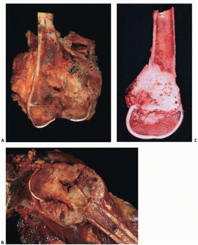
FIGURE 9.17. Osteosarcoma (gross). Distal femur (A), proximal humerus (B), and distal femur (C) with sclerotic lesions of the metaphysis destroying cortical bone and extending into soft tissue. Growth plate is a relative barrier. Color varies from ivory-white to yellow to red, depending on the amount of bone, necrosis, and hemorrhage.

Prognostic indicators include the size and extent of cortical and soft tissue penetration and weight loss of greater than 10 pounds.


  • With the advent of neoadjuvant chemotherapy and surgical removal, the 5- and 10-year survivals of children with osteogenic sarcoma without evidence of disseminated disease at diagnosis is considerably higher and is in the 70 percent to 80 percent range.

John Healey, at Memorial Sloan-Kettering Center, has reported 80 percent survival rates for Stage IIB lower limb lesions, and 90 percent for Stage IIA. Tibial lesions had a better survival than distal femoral lesions. He found metastases a bad prognostic factor, with only 15 percent of patients with distant metastases cured (51).

In recent years, the standard treatment has evolved into chemotherapeutic regimens consisting predominantly of


  • doxorubicin,


  • cisplatin, and


  • high-dose metotrexate.


FIGURE 9.18. Osteosarcoma (gross). Codman triangle. Distal femur with tumor raising the periosteum. (A) Low power and specimen x-ray. (B) Higher power. Borders of Codman triangle are the bone surface, the tumor, and the periosteal surface.


FIGURE 9.19. Osteosarcoma (histopathology). Bone marrow is totally replaced by tumor (A), which, at high power, reveals pleomorphic, mitotic cells making malignant osteoid (B). Osteosarcomas demonstrate a wide variety of histologic features, with fibrous and chondroid areas suggesting a painter’s palette (C, D). (Continued)

Only gold members can continue reading. Log In or Register to continue

Stay updated, free articles. Join our Telegram channel

Jul 24, 2016 | Posted by in PATHOLOGY & LABORATORY MEDICINE | Comments Off on Bone-Forming Bone Tumors and Tumor-like Lesions

Full access? Get Clinical Tree

Get Clinical Tree app for offline access