(A) Decreased parathyroid hormone
(B) Increased amount of woven bone
(C) Osteoclastic bone tunneling or dissecting bone resorption
(D) Peritrabecular marrow fibrosis
(E) Subperiosteal resorption on the radial side of middle phalanges
23. Which of the following conditions is associated with dystrophic calcification?
(A) Hyperparathyroidism
(B) Infarction
(C) Metastatic disease
(D) Myeloma
(E) Sarcoidosis
24. Which of the following pathologic features is not associated with osteoarthritis?
(A) Eburnated articular surface
(B) Osteophyte formation
(C) Rice bodies
(D) Subchondral cyst formation
(E) Surface fibrillation
25. A 25-year-old man presents with toe pain, and is found to have a lobulated, ossified mass in the soft tissue that does not seem to be attached to the distal phalanx. Histologically, the lesion is composed of lobulated, cellular cartilaginous tissue, with calcified fibrocartilagenous tissue toward’s the periphery. What is the best diagnosis for this lesion?
(A) Florid reactive periostitis
(B) Nora’s lesion
(C) Osteochondroma
(D) Osteoma
(E) Subungual exostosis
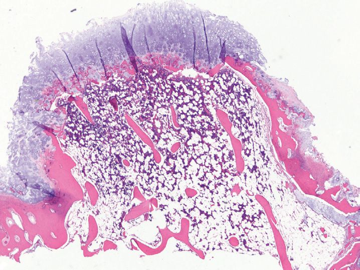
26. Which of the following clinicopathologic features is least likely to be associated with this painful lesion excised from the left leg of a 65-year-old man (see Figure 5-2)?
(A) Contains a central nidus
(B) Immature woven bone forms the major component of the lesion
(C) More than 50% cases occur in the lower extremities
(D) Painless lesion
(E) Usually measures <1 cm
27. A 40-year-old man is found to have an incidental, solitary, homogeneously dense intramedullary lesion in the left pelvic bone. The lesion has spiculated margins that blend with the adjacent cancellous bone. Histologically, it is composed entirely of lamellar bone with very minimal woven bone. What is the best diagnosis?
(A) Enostosis (bone island)
(B) Intramedullary osteosarcoma
(C) Osteoblastic metastasis
(D) Osteoblastoma
(E) Osteoid osteoma
28. Which cortical surface-based bone-forming tumor grossly mimics a normal bone, and when multifocal, is associated with Gardner’s syndrome?
(A) Conventional osteosarcoma
(B) Low-grade surface osteosarcoma
(C) Osteoblastoma
(D) Osteoid osteoma
(E) Osteoma
29. Which of the listed features favors a diagnosis of osteosarcoma over aggressive osteoblastoma?
(A) Coarse lace-like osteoid deposition
(B) Distinct osteoblastic rimming
(C) Epithelioid osteoblasts
(D) Infiltrative borders
(E) Lack of atypical mitoses
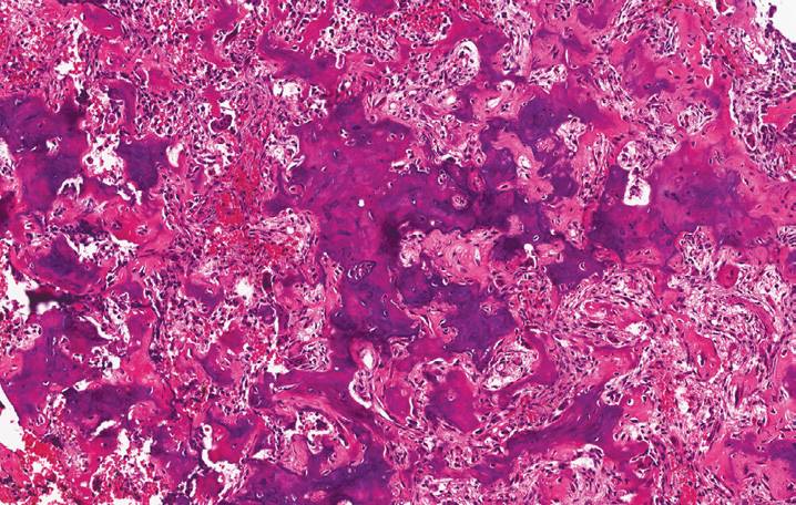
30. Which of the following clinicopathologic features is least likely to be associated with the bone tumor shown in Figure 5-3?
(A) A minimum of 10% malignant osteoid production is required for a diagnosis
(B) Bimodal age distribution
(C) Metaphysis is the most common location
(D) More common in males than in females
(E) Most common primary non-hematopoeitic malignancy of the skeletal system
31. Which of the following subtypes is the least common subtype of osteosarcoma?
(A) Conventional high-grade osteosarcoma
(B) High-grade secondary osteosarcoma
(C) Intramedullary well-differentiated osteosarcoma
(D) Parosteal osteosarcoma
(E) Periosteal osteosarcoma
32. Which of the following features is helpful in classifying a surface osteosarcoma as a periosteal osteosarcoma as opposed to a parosteal osteosarcoma?
(A) Broad-based, well-demarcated, solid, tan-white lesion
(B) Cellular spindle cell proliferation with limited cytologic atypia
(C) Minimal periosteal reaction on radiologic examination
(D) Neoplastic cartilaginous areas admixed with severe cytologic atypia
(E) Tumor located in the posterior aspect of proximal femur
33. Which of the following clinical conditions/syndromes is not considered to be a risk factor for the development of osteosarcoma?
(A) Bilateral retinoblastomas
(B) Chemotherapy
(C) Chronic osteomyelitis
(D) Li-Fraumeni syndrome
(E) Paget’s disease
34. Which of the following tumor is commonly associated with the formation of Codman triangle?
(A) Aneurysmal bone cyst
(B) Chondrosarcoma
(C) Ewing sarcoma
(D) Fibrous dysplasia
(E) Osteogenic sarcoma
35. Which of the following cartilage-forming tumors is the best diagnosis for the lesion shown in this image? (see Figure 5-4)
(A) Chondroblastoma
(B) Chondrosarcoma
(C) Enchondroma
(D) Osteochondroma
(E) Periosteal chondroma
36. A well-demarcated lytic lesion encountered in the epiphysis of a 20-year-old man is shown in Figure 5-5. Which of the following features is not typically related to this lesion?
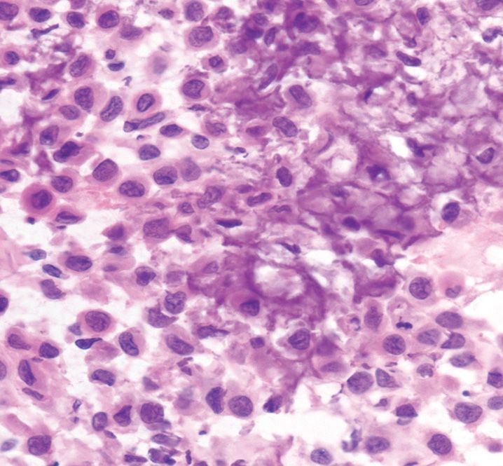
(A) Abundant blue hyaline cartilage
(B) Chicken wire calcifications
(C) Irregular distribution of osteoclastic giant cells
(D) Nuclear grooves
(E) Secondary aneurysmal bone cyst formation
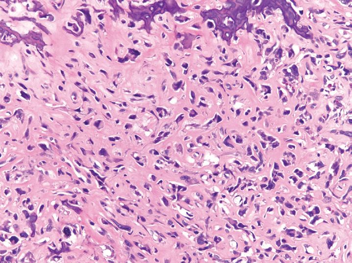
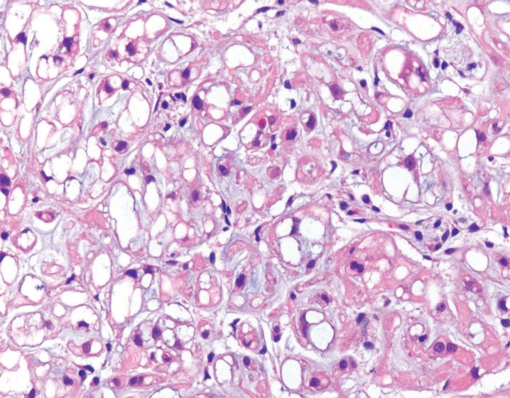
37. An intramedullary tumor resected from a 35-year-old man is shown in Figure 5-6. All the following features are associated with this tumor, except
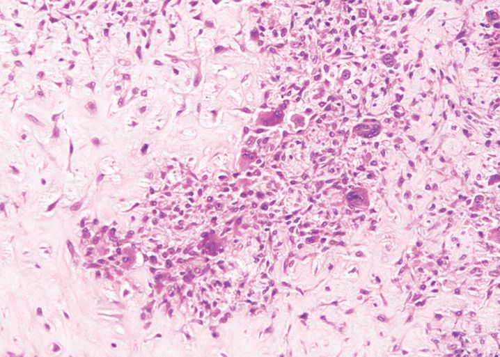
(A) Increased cellularity toward the periphery of the lobules
(B) Minimal cytologic atypia
(C) Nodules of hyaline cartilage
(D) Osteoclast-like giant cells
(E) Spindle or stellate cells with long cytoplasmic processes
38. Which of the following radiologic/pathologic feature, when present in a well-differentiated cartilage-forming lesion, warrants a diagnosis of a grade 1 chondrosarcoma?
(A) Dark nuclei with details visible at 400 × magnification
(B) Islands of cartilage permeating viable, lamellar bone
(C) Low cellularity with round chondrocytes within lacunae
(D) Ring calcifications
(E) Slow-growing mass
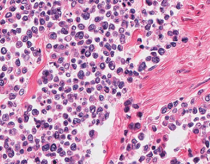
39. Based on the histologic features shown in Figure 5-7, which of these findings would be unusual for this lesion?
(A) Commonly occurs in the ribs, jaw, and pelvis
(B) Gritty calcifications, cystic change, hemorrhage, and necrosis, are common
(C) Hemangiopericytoma-like vascular pattern
(D) Peak age of occurrence is 70 years
(E) The smaller cells may express CD99
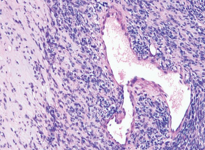
40. A 19-year-old teenager undergoes radiologic evaluation for knee pain and is found to have a 4.5-cm lytic lesion involving the metaphysis of the distal femur. It appears to be eccentrically located within the medullary cavity and is juxtaposed to the cortex. Based on the histologic findings shown in Figure 5-8, what is the best diagnosis?
(A) Benign fibrous histiocytoma
(B) Desmoplastic fibroma
(C) Fibrous dysplasia
(D) Giant cell tumor
(E) Nonossifying fibroma
41. An 8-year-old boy presents with a tumor involving the diaphysis of femur (see Figure 5-9). Imaging studies reveal the presence of “onion skinning” and “sunburst” pattern of periosteal reaction. Which of the following genes is most frequently involved in the reciprocal translocation diagnostic of this tumor?
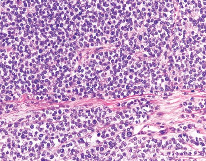
(A) E1AF, chromosome 2
(B) ERG, chromosome 21
(C) ETV1, chromosome 7
(D) ETV4, chromosome 17
(E) FLI1, chromosome 11
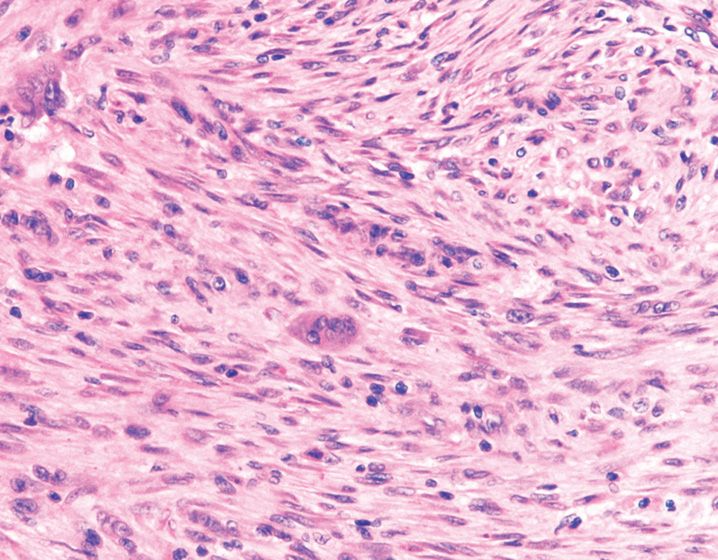
42. A 23-year-old woman undergoes curettage of a lytic lesion located in the distal femur. Which of the following features is not characteristic of this tumor? (See Figure 5-10.)
(A) Less than 2% of tumors may metastasize to the lung
(B) May show foci of osteoid deposition
(C) Nuclei of multinucleated giant cells are different from those of the mononuclear cell population
(D) Numerous mitotic figures may be found
(E) Tumor may demonstrate destruction of the cortex
43. A 60-year-old man presents with low back pain and is diagnosed with this sacrococcygeal tumor (see Figure 5-11). Which of the following immunohisto-chemical stains is not expressed by this tumor?
(A) Brachyury
(B) CDX-2
(C) Cytokeratin 8/18
(D) Epithelial membrane antigen (EMA)
(E) S-100
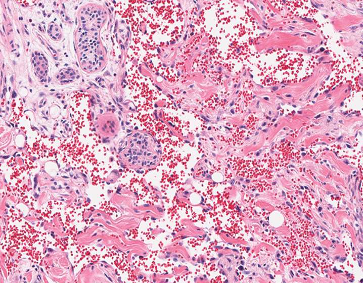
44. A 30-year-old man with history of trauma is found to have an expansile lytic lesion in the midshaft of right tibia. Imaging shows a “soap-bubble” appearance. Based on the image provided (see Figure 5-12), what is the best diagnosis?
(A) Adamantinoma
(B) Benign fibrous histiocytoma
(C) Fibrous dysplasia
(D) Giant cell tumor
(E) Nonossifying fibroma
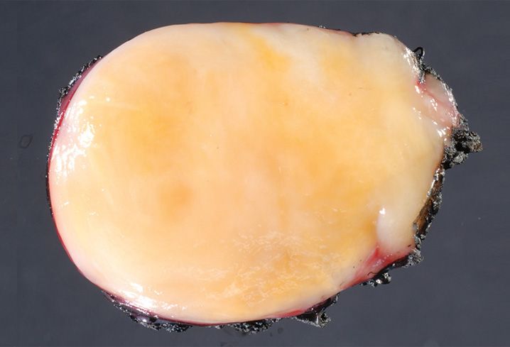
45. A rib tumor with sclerotic rind-like periphery and ground glass appearance resected from a 35-year-old man is shown in Figure 5-13. What is the best diagnosis for this lesion?
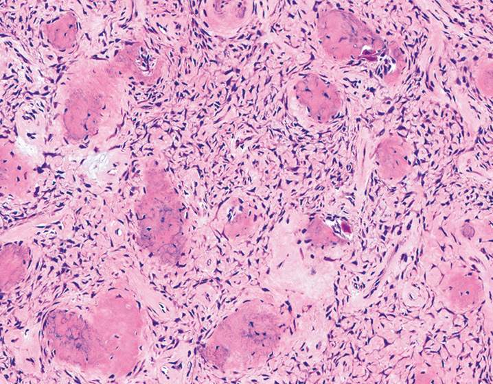
(A) Cemento-ossifying fibroma
(B) Desmoplastic fibroma
(C) Fibrous dysplasia
(D) Nonossifying fibroma
(E) Osteofibrous dysplasia
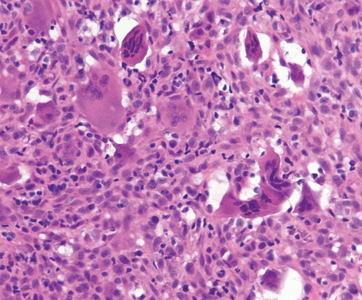
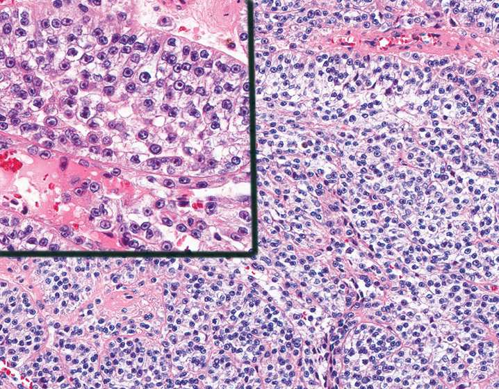
46. A 12-year-old boy undergoes resection of a rapidly growing forearm mass shown in Figure 5-14. Which of the following features is least likely to be associated with this lesion?
(A) Atypical mitotic activity can be seen
(B) Lesional cells are diffusely positive for smooth muscle actin
(C) Often contains extravasated red blood cells
(D) Often located in the deep subcutis
(E) Osteoclast-like giant cells are present in 10% of cases
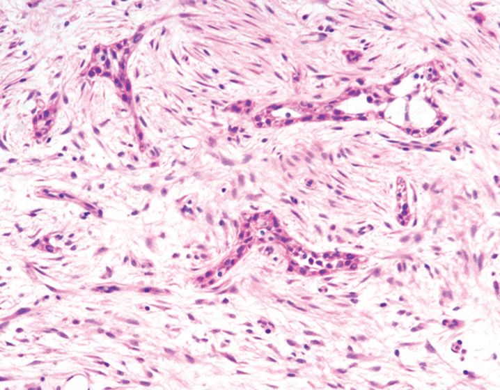
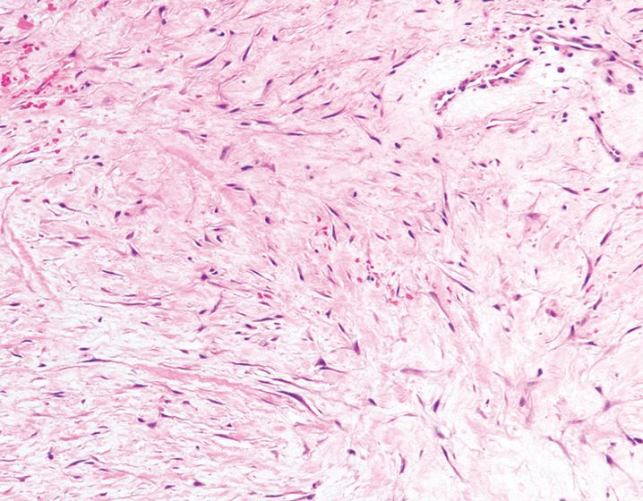
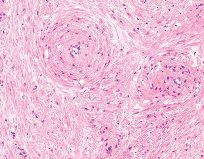
47. A 60-year-old woman undergoes resection of a soft tissue mass located in the lower scapula (see Figure 5-15). What is the best diagnosis for this lesion?
(A) Desmoplastic fibroblastoma
(B) Elastofibroma
(C) Fibroma of tendon sheath
(D) Keloid
(E) Spindle cell lipoma
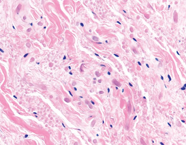
48. What is the best diagnosis for this superficially located axillary mass excised from a 2-year-old infant (see Figure 5-16)?
(A) Calcifying aponeurotic fibroma
(B) Fibrous hamartoma of infancy
(C) Giant cell fibroblastoma
(D) Myofibroma
(E) Lipofibromatosis
49. What percentage of cases of desmoid-type fibromatosis harbor mutations in the β-catenin gene ?
(A) 10%
(B) 15%
(C) 25%
(D) 50%
(E) 90%
50. A 45-year-old woman is diagnosed with myxoinflammatory fibroblastic sarcoma. Which of the following features is not typical of this tumor?
(A) Hyalinized and myxoid areas
(B) Lymphocytes and plasma cell-rich inflammation
(C) Pseudolipoblast-like cells
(D) Reed-Sternberg like cells
(E) Well-developed arborizing thick-walled vasculature
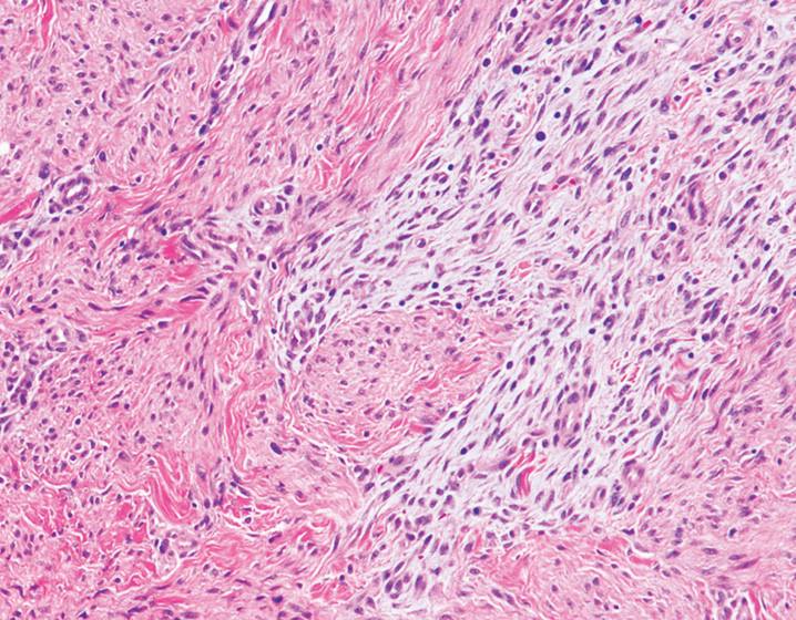
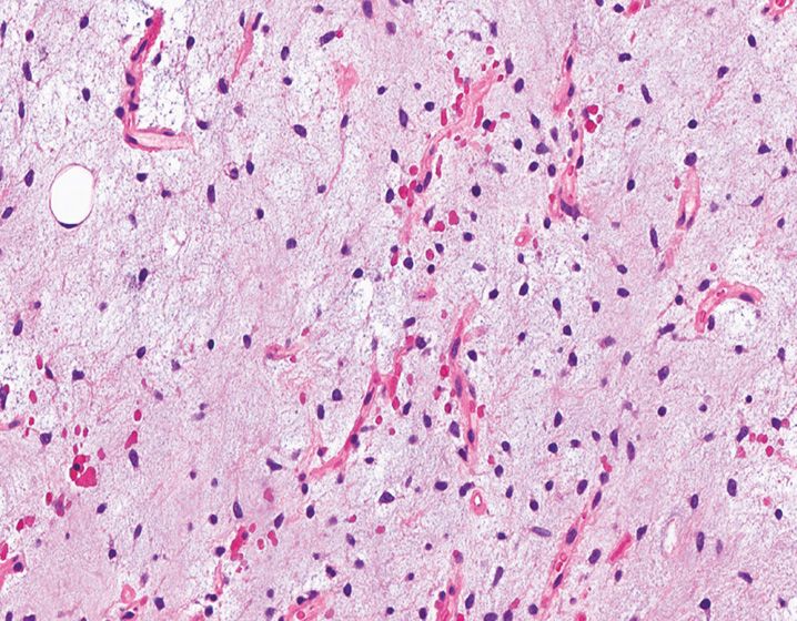
51. A 35-year-old man undergoes resection of this tumor located in upper thigh (see Figure 5-17). Which of the following molecular abnormalities is specific for this tumor?
(A) t(2;13)
(B) t(7;16)
(C) t(12;15)
(D) t(12;22)
(E) t(17;22)
52. A 70-year-old man with a 1.5-cm superficial neck lesion is diagnosed with atypical fibroxanthoma. Which of the following features is associated with this tumor?
(A) Overlying epidermis is usually hyperplastic
(B) Patients have hyperlipidemia
(C) The lesional cells express CD68 and desmin
(D) Touton-type giant cells are diagnostic of this entity
(E) Usually occurs in sun-exposed areas
53. A 3.5-cm nodule excised from the trunk of a 40-year-old man shows a storiform arrangement of cells, with infiltration into the subcutaneous tissue in a honeycomb pattern. Which of immunohistochemical markers would be positive in the tumor cells?
(A) Caldesmon
(B) CD34
(C) Desmin
(D) S-100
(E) Smooth muscle actin
54. Angiomatoid fibrous histiocytoma is least likely to be associated with which of the following features?
(A) Cystic change
(B) Lesional cells that are positive for CD31 and CD34
(C) Occurs in children and young adults
(D) Shows molecular alterations similar to clear cell sarcoma of soft tissue
(E) Shows peripheral fibrous capsule with lymphocytic inflammation
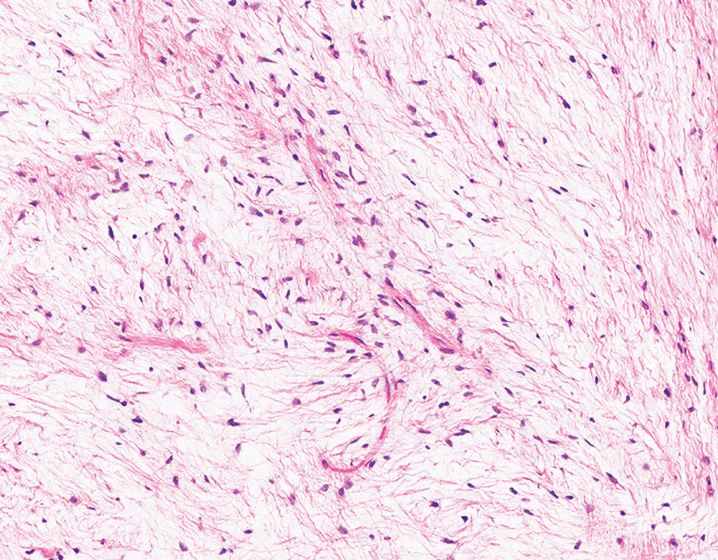
55. A 52-year-old man undergoes excision of this poorly circumscribed mass located in the posterior aspect of neck (see Figure 5-18). What is the best diagnosis?
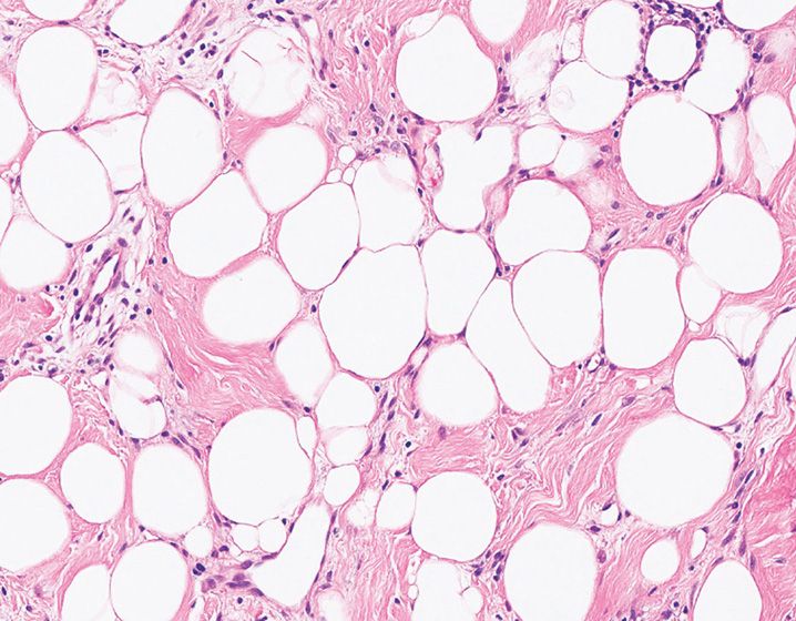
(A) Angiolipoma
(B) Elastofibroma
(C) Intramuscular lipoma
(D) Liposarcoma
(E) Spindle cell lipoma
56. Which of the following adipocytic lesions does not contain lipoblasts?
(A) Angiolipoma
(B) Chondroid lipoma
(C) Lipoblastoma
(D) Liposarcoma
(E) Pleomorphic lipoma
57. All the listed features are characteristic of a dedifferentiated liposarcoma, except
(A) Behaves less aggressively than other high-grade pleomorphic sarcomas
(B) Can recur entirely as a well-differentiated liposarcoma
(C) Dedifferentiated foci have to show high-grade morphology
(D) Grossly presents as a distinct tan-white firm nodule in a background of adipose tissue
(E) Retroperitoneum is the most common location
58. Which of the following genes is implicated in the karyotypic aberration found in this thigh mass resected from a 40-year-old man (see Figure 5-19)?
(A) CDK4
(B) DDIT3
(C) HMGA2
(D) MDM2
(E) OS1
59. A 52-year-old woman undergoes excision of a left leg nodule that is shown in Figure 5-20. Which of the following clinicopathologic features is not typical of this lesion?
(A) Degenerative atypia
(B) Diffuse immunoreactivity with h-caldesmon
(C) Extremities are commonly involved
(D) Pain is a common presenting symptom
(E) Venous subtype is most common histologic subtype
60. A 16-year-old teenager with history of renal transplant develops multiple liver nodules. A biopsy of one of these nodules shows Epstein–Barr virus (EBV)-associated smooth muscle tumor. Which of the following features is least likely to be associated with this entity?
(A) Composed of well-differentiated and small, primitive smooth muscle cells
(B) Lung and liver are commonly involved
(C) Lymphocytic inflammatory infiltrate
(D) Marked cytologic atypia
(E) Usually arises approximately 3 years post-transplantation
61. A 4-year-old girl was found to have a polypoid soft tissue mass composed of bland spindle cells and myotubules arranged in a myxoid background. The lesion also shows occasional strap cells. What is the best classification for this lesion?
(A) Adult rhabdomyoma
(B) Botyroid rhabdomyosarcoma
(C) Cellular rhabdomyoma
(D) Fetal rhabdomyoma
(E) Genital rhabdomyoma
62. A 7-year-old girl undergoes resection of a cervical polypoid tumor composed of small, undifferentiated spindle-to-epithelioid cells that are tightly packed in a band-like arrangement beneath the epithelial surface. The cells are diffusely positive for MyoD1. What is the best diagnosis?
(A) Anaplastic rhabdomyosarcoma
(B) Alveolar rhabdomyosarcoma
(C) Botyroid rhabdomyosarcoma
(D) Embryonal rhabdomyosarcoma
(E) Spindle cell rhabdomyosarcoma
63. A 20-year-old man undergoes resection of a thigh mass shown in Figure 5-21. This lesion is diffusely positive for myogenin and negative for cytokeratin. Which of the following findings is not associated with this entity?
(A) Intracytoplasmic PAS-positive crystalline inclusions
(B) Multinucleated giant cells
(C) PAX7/FKHR translocation
(D) Poor clinical prognosis
(E) Rapidly growing mass
64. Which of the following vascular proliferations commonly occurs on the thumb, and shows an intravascular proliferation of bland endothelial cells within a thrombus?
(A) Bacillary angiomatosis
(B) Capillary hemangioma
(C) Glomeruloid hemangioma
(D) Spindle cell hemangioma
(E) Papillary endothelial hyperplasia
65. Bacillary angiomatosis is associated with all of the following features except
(A) Affects immunocompromised individuals
(B) Caused by infection with Human Herpes Virus-8
(C) Immunoreactivity with FLI-1 antibody
(D) Neutrophils and eosinophilic fibrin aggregates
(E) Visceral involvement is in the form of peliosis
66. Which of the following vascular lesions is associated with multisystem disease characterized by polyneuropathy, organomegaly, endocrinopathy, monoclonal gammopathy, and skin changes (POEMS syndrome)?
(A) Bacillary angiomatosis
(B) Capillary hemangioma
(C) Glomeruloid hemangioma
(D) Spindle cell hemangioma
(E) Papillary endothelial hyperplasia
67. Which of the following immunohistochemical markers is specifically expressed by juvenile capillary hemangioma and not by other vascular tumors?
(A) CD31
(B) CD34
(C) FLI-1
(D) GLUT1
(E) vWF
68. Which of the following vascular tumors is frequently found in patients with Maffucci syndrome and mimics Kaposi’s sarcoma?
(A) Bacillary angiomatosis
(B) Capillary hemangioma
(C) Glomeruloid hemangioma
(D) Spindle cell hemangioma
(E) Papillary endothelial hyperplasia
69. Which of the following vascular lesions shows a central damaged vessel, an eosinophil-rich inflammatory infiltrate, and a characteristic “tombstone” pattern of endothelial cells?
(A) Deep hemangioma
(B) Epithelioid hemangioendothelioma
(C) Epithelioid hemangioma
(D) Glomeruloid hemangioma
(E) Kaposiform hemangioendothelioma
70. A 75-year-old man undergoes resection of this vascular tumor located in the liver. Which of the following markers is the least sensitive immunohistochemical marker of this lesion? (See Figure 5-22.)
(A) CD31
(B) CD34
(C) ERG
(D) FLI1
(E) vWF
71. Which of the following parts of a nerve does neurofibroma arise from?
(A) Axon
(B) Endoneurium
(C) Fibroblasts
(D) Perineurium
(E) Schwann cell
72. Which of the following peripheral nerve sheath tumors has the highest risk for malignant transformation?
(A) Cutaneous NF1-associated neurofibroma
(B) Deep NF1-associated neurofibroma
(C) Non-NF1-associated neurofibroma
(D) Plexiform neurofibroma
(E) Solitary neurofibroma
73. Which of the following morphologic features is considered to be the diagnostic hallmark of diffuse neurofibroma?
(A) Hyalinization
(B) Multiple intraneural tumor nodules
(C) Myxoid change
(D) Wagner–Meissner bodies
(E) Storiform arrangement of cells
74. Which of the following findings is not associated with a neurofibroma undergoing malignant transformation?
(A) Cytologic atypia
(B) Increased cellularity
(C) Mitotic activity
(D) Necrosis
(E) Prominent S-100 immunoreactivity
75. Figure 5-23 shows a thigh mass that was resected from the peripheral aspect of a large nerve. Which of the following findings is not associated with this lesion?
(A) Admixture of S-100, CD34, and EMA-positive cells
(B) Encapsulation
(C) Myxoid areas
(D) Perivascular hyalinization
(E) Verocay bodies
76. Which of the following clinical syndromes/complexes is observed in up to 50% of patients with psammomatous melanotic schwannoma?
(A) Carney complex
(B) Klippel-Trenaunay syndrome
(C) MEN-2
(D) NF1
(E) NF2
77. A 60-year-old man undergoes resection of a buttock mass composed of malignant spindle cells with geographic areas of necrosis and perivascular accentuation. They are weakly positive for S-100 and negative for cytokeratin. What is the best diagnosis?
(A) Fibrosarcoma
(B) Leiomyosarcoma
(C) Malignant peripheral nerve sheath tumor
(D) Malignant solitary fibrous tumor
(E) Monophasic synovial sarcoma
78. In a patient diagnosed with neuroblastoma, which of the following factors is associated with a good clinical prognosis?
(A) Age at diagnosis <1 year
(B) Deletion of chromosome 1p36
(C) Higher stage
(D) High mitosis–karyorrhexis index
(E) Necrosis
79. A soft tissue mass resected from a 20-year-old man is shown in Figure 5-24. Molecular studies are positive for t(12;22). Which of these locations is the most common site of involvement by this tumor?
Stay updated, free articles. Join our Telegram channel

Full access? Get Clinical Tree


