TECHNIQUE |
FINDINGS |
|---|
PERIPHERAL ARTERIES |
Palpate arterial pulses in neck and extremities |
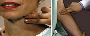
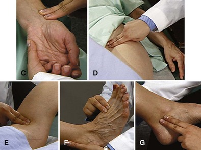
Palpate carotid, brachial, radial, femoral, popliteal, dorsalis pedis, and posterior tibial arteries, using distal pads of second and third fingers, as shown in figures below and on p. 125. |
|
|
 Characteristics Characteristics
Compare characteristics bilaterally, as well as between upper and lower extremities. |
EXPECTED:Femoral pulse as strong as or stronger than radial pulse. |
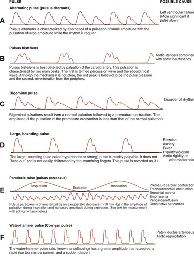
UNEXPECTED:Femoral pulse weaker than radial pulse or absent. Alternating pulse (pulsus alternans), pulsus bisferiens, bigeminal pulse (pulsus bigeminus), bounding pulse, labile pulse, paradoxical pulse (pulsus paradoxus), pulsus differens, tachycardia, trigeminal pulse (pulsus trigeminus), or water-hammer pulse (Corrigan pulse). |
|
 Rate Rate |
EXPECTED:60 to 90 beats per minute. |
UNEXPECTED:Rate different from that observed during cardiac examination. |
 Rhythm Rhythm |
EXPECTED:Regular. |
UNEXPECTED:Irregular, either in a pattern or patternless. |
 Contour Contour |
EXPECTED:Smooth, rounded, or dome shaped. |
 Amplitude Amplitude |
UNEXPECTED:Bounding, full, diminished, or absent. Describe on scale of 0 to 4:
|
Auscultate temporal, carotid, and subclavian arteries; abdominal aorta; and renal, iliac, and femoral arteries for bruits |
When auscultating the carotid vessels, you may at times need to ask patient to hold breath for a few heartbeats. Auscultate with bell of stethoscope. |
UNEXPECTED:Transmitted murmurs, bruits. |
Assess for arterial occlusion and insufficiency |
 Site Site
Assess for pain distal to possible occlusion. |
UNEXPECTED:Dull ache accompanied by fatigue and often crampiness; possible constant or excruciating pain. Weak, thready, or absent pulses; systolic bruits over arteries; loss of body warmth; localized pallor or cyanosis; delay in venous filling; or thin, atrophied skin, muscle atrophy, and loss of hair. |
 Degree of occlusion Degree of occlusion
Ask patient to lie supine.
Elevate extremity, note degree of blanching, then ask patient to sit on edge of table or bed to lower extremity. Note time for maximal return of color when extremity is lowered. |
EXPECTED:Slight pallor on elevation and return to full color as soon as leg becomes dependent. |
UNEXPECTED:Delay of more than 2 seconds. |
Measure blood pressure |
Measure in both arms at least once. Patient’s arm should be slightly flexed and comfortably supported on table, pillow, or your hand. |
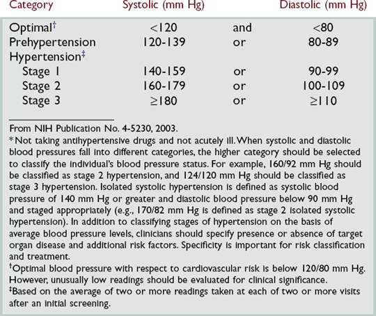
EXPECTED:100 to 140 mm Hg systolic and 60 to 90 mm Hg diastolic, with pulse pressure of 30 to 40 mm Hg (sometimes to 50 mm Hg). Reading between arms may vary by as much as 10 mm Hg; usually higher in right arm. Prehypertension is now defined as a blood pressure between 120 and 139 mm Hg systolic or 80 and 89 mm Hg diastolic. |
UNEXPECTED:Hypertension (see table below). |