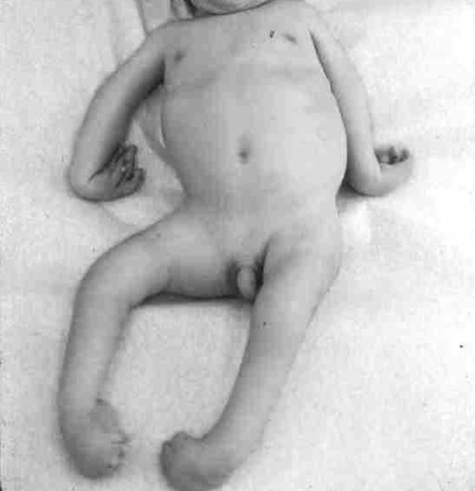
In contrast to malformations, deformations are caused by extrinsic factors impinging physically on the fetus during development. They are especially common during the second trimester of development when the fetus is constrained within the amniotic sac and uterus. For example, contractions of the joints of the extremities, known as arthrogryposes, in combination with deformation of the developing skull, occasionally accompany constraint of the fetus due to twin or triplet gestations or prolonged leakage of amniotic fluid (Fig. 14-2). Most deformations apparent at birth either resolve spontaneously or can be treated by external fixation devices to reverse the effects of the instigating cause.
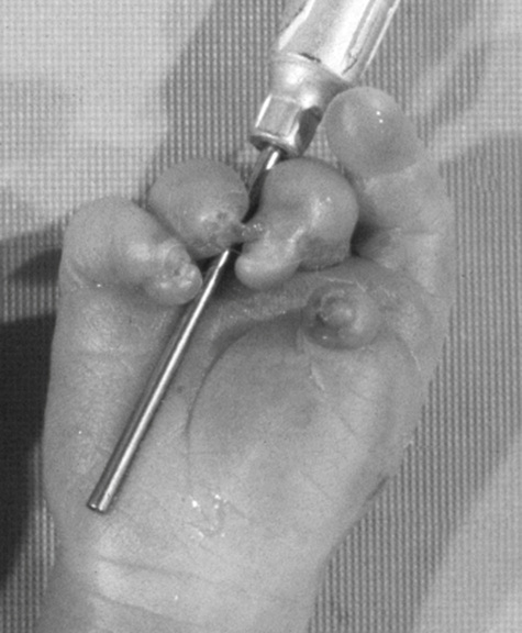
Disruptions, the third category of birth defect, result from destruction of irreplaceable normal fetal tissue. Disruptions are more difficult to treat than deformations because they involve actual loss of normal tissue. Disruptions may be the result of vascular insufficiency, trauma, or teratogens. One example is amnion disruption, the partial amputation of a fetal limb associated with strands of amniotic tissue. Amnion disruption is often recognized clinically by the presence of partial and irregular digit amputations in conjunction with constriction rings (Fig. 14-3).
The pathophysiological concepts of malformations, deformations, and disruptions are useful clinical guides to the recognition, diagnosis, and treatment of birth defects, but they sometimes overlap. For example, vascular malformations may lead to disruption of distal structures, and urogenital malformations that cause oligohydramnios can cause fetal deformations. Thus a given constellation of birth defects in an individual may represent combinations of malformations, deformations, and disruptions.
Genetic, Genomic, and Environmental Causes of Malformations
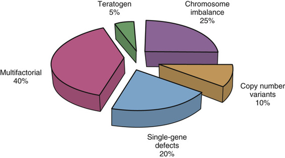
Malformations have many causes (Fig. 14-4). Chromosome imbalance accounts for approximately 25%, of which autosomal trisomies for chromosomes 21, 18, and 13 (see Chapter 6) are some of the most common. The recent clinical application of genome-wide arrays in comparative genomic hybridization (CGH or array-CGH; see Chapter 5) has revealed small, de novo submicroscopic deletions and/or duplications, also known as copy number variants (CNVs), in as many as 10% of individuals with birth defects. An additional 20% are caused by mutations in single genes. Some malformations, such as achondroplasia or Waardenburg syndrome, are inherited as autosomal dominant traits. Many heterozygotes with birth defects, however, represent new mutations that are so severe that they are genetic lethals and are therefore often found to be isolated cases within families (see Chapter 7). Other malformation syndromes are inherited in an autosomal or X-linked recessive pattern, such as the Smith-Lemli-Opitz syndrome or the Lowe syndrome, respectively.
Stay updated, free articles. Join our Telegram channel

Full access? Get Clinical Tree


