8 Biliary Diseases
Anatomy of the Extrahepatic Biliary System
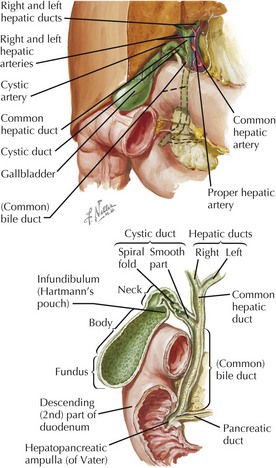
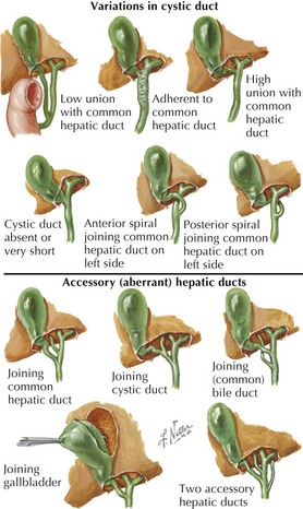
• Anatomy of the biliary system is highly variable, and this includes ducts, arteries, veins, and lymphatics.
Gallbladder
• Normally lies between hepatic segments IV and V, in a ventral fossa between the anatomical right and left lobes


• Parasympathetic preganglionic innervation from left (anterior) vagus fibers contracts gallbladder and relaxes bile duct sphincter.
• Postganglionic sympathetic fibers from the celiac ganglion are driven by preganglionic fibers from T7-T10 spinal segments traveling in greater splanchnic nerves.
Cystic Duct
• Typical cystic duct joins the common hepatic duct well below the right and left hepatic duct junction.
• Triangle of Calot: classic configuration (shown above) with cystic duct right, common bile duct left, liver above, and right hepatic artery passing through


(Common) Bile Duct
• Bile duct sphincter: smooth muscle surrounding the distal end of the duct, part of the complex sphincter of Oddi


