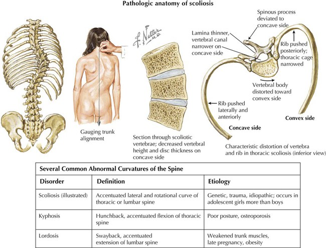
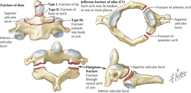
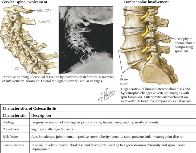
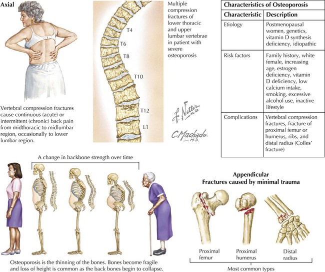
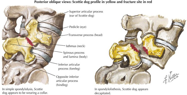
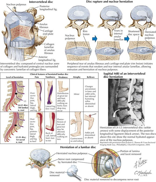
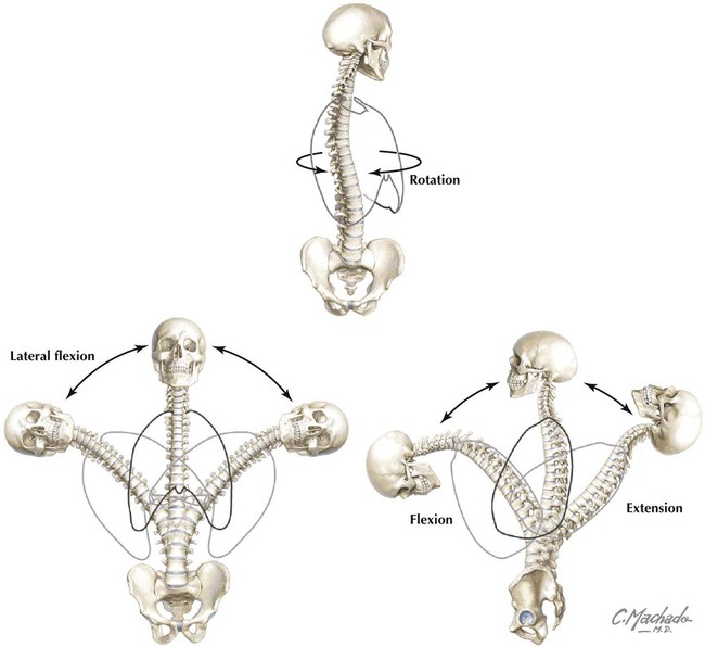
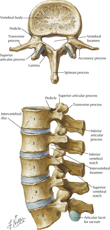
• Support. The vertebral column forms the axis of the body and is critical for upright posture (standing or sitting), as a support for the head, as an attachment point and brace for movements of the upper limbs, and as a support for transferring the weight of the trunk to the lower limbs. • Protection. The vertebral column protects the spinal cord and proximal portions of the spinal nerves before they distribute throughout the body. • Movements. Muscles of the back function in movements of the head and upper limbs and in support and movements of the vertebral column. Figure 2-1 shows key surface landmarks of the back, including the following bony landmarks: • Vertebrae prominens: the spinous process of the C7 vertebra, usually the most prominent process in the midline at the posterior base of the neck. • Scapula: part of the pectoral girdle that supports the upper limb; note its spine, inferior angle, and medial border. • Iliac crests: felt best when you place your hands “on your hips.” An imaginary horizontal line connecting the iliac crests passes through the spinous process of the vertebra L4 and the intervertebral disc of L4-L5, providing a useful landmark for lumbar puncture or epidural block (see Clinical Focus 2-11). • Posterior superior iliac spines: an imaginary horizontal line connecting these two points passes through the spinous process of S2 (second sacral segment). The vertebral column (spine) forms the central axis of the human body, highlighting the segmental nature of all vertebrates, and usually is composed of 33 vertebrae distributed as follows (Fig. 2-2): • Cervical: seven total; first two called the atlas (C1) and axis (C2). • Thoracic: 12 total; each articulates with a pair of ribs. • Lumbar: five total; large vertebrae for support of the body’s weight. • Sacral: five fused vertebrae for stability in the transfer of weight from the trunk to the lower limbs. • Coccyx: four total; Co1 often is not fused, but Co2-Co4 are fused (a remnant of embryonic tail). The actual number of vertebrae can vary, especially the number of coccygeal vertebrae. Viewed from the lateral aspect (Fig. 2-2), one can identify the following: • Cervical curvature (cervical lordosis): a secondary curvature acquired when the infant can support the weight of the head. • Thoracic curvature (thoracic kyphosis): a primary curvature present in the fetus (imagine the spine in the “fetal position”). • Lumbar curvature (lumbar lordosis): a secondary curvature acquired when the infant assumes an upright posture and supports its own weight. • Sacral curvature: a primary curvature present in the fetus. A “typical” vertebra has the following features (Fig. 2-3): • Arch: a projection formed by paired pedicles and laminae. • Articular processes (facets): two superior and two inferior facets for articulation with adjacent vertebrae. • Body: the weight-bearing portion of a vertebra that tends to increase in size as one descends the spine. • Intervertebral foramen (foramina): the opening formed by the vertebral notches that is traversed by spinal nerve roots and associated vessels. • Lamina (laminae): paired portions of the vertebral arch that connect the transverse processes to the spinous process. • Pedicle: paired portions of the vertebral arch that attach the transverse processes to the body. • Transverse foramina: apertures that exist in transverse processes of cervical vertebrae only and transmit the vertebral vessels. • Transverse processes: the lateral extensions from the union of the pedicle and lamina. • Spinous process: a projection that extends posteriorly from the union of two laminae. • Vertebral foramen (canal): a foramen formed from the vertebral arch and body that contains the spinal cord and its meningeal coverings. • Vertebral notches: superior and inferior semicircular features that in articulated vertebrae form an intervertebral foramen (two semicircular notches form a circle). The cervical spine is composed of seven cervical vertebrae. The first two cervical vertebrae are unique and called the atlas and axis (Fig. 2-4). The atlas (C1) holds the head on the neck (the titan Atlas of Greek mythology held the heavens on his shoulders as punishment by Zeus). The axis (C2) is the point of articulation where the head turns on the neck, providing an “axis of rotation.” Table 2-1 summarizes key features of the cervical vertebrae. The cervical region is a fairly mobile portion of the spine, allowing for flexion and extension as well as rotation and lateral bending. TABLE 2-1 Key Features of the Cervical Vertebrae (C1-C7) The thoracic spine is composed of 12 thoracic vertebrae (Fig. 2-5 and Table 2-2). The 12 pairs of ribs articulate with the thoracic vertebrae. This region of the spine is more rigid and inflexible than the cervical region. TABLE 2-2 Key Features of Thoracic, Lumbar, Sacral, and Coccygeal Vertebrae The lumbar spine is composed of five lumbar vertebrae (see Figs. 2-3 and 2-5 and Table 2-2). The lumbar vertebrae are comparatively large for bearing the weight of the trunk and are fairly mobile, but not nearly as mobile as the cervical vertebrae. The sacrum is composed of five fused vertebrae that form a single, wedge-shaped bone (Fig. 2-5 and Table 2-2). The sacrum provides support for the pelvis. The coccyx is a remnant of the embryonic tail and usually consists of four vertebrae, with the last three often fused into a single bone. The coccyx lacks vertebral arches and has no vertebral canal. The craniovertebral joints include the atlanto-occipital (atlas and occipital bone of the skull) and atlanto-axial (atlas and axis) joints. Both are synovial joints that provide a relatively wide range of motion compared with other joints of the vertebral column. The atlanto-occipital joint permits one to nod the head up and down (flexion and extension), whereas the atlanto-axial joint is a pivot joint that permits one to rotate the head from side to side, as if to indicate “no” (Fig. 2-6 and Table 2-3). TABLE 2-3 Key Features of Atlanto-occipital and Atlanto-axial Joints The joints of the vertebral arches (zygapophysial joints) occur between the superior and inferior articular processes (facets) of adjacent vertebrae and allow for some gliding or sliding movement (Fig. 2-7 and Table 2-4). These joints slope inferiorly in the cervical spine (facilitate flexion and extension), are more vertically oriented in the thoracic region (limit flexion and extension but allow for rotation), and are interlocking in the lumbar spine (but do allow flexion and extension, but not to the degree present in the cervical spine). Corresponding ligaments connect the spinous processes, laminae, and bodies of adjacent vertebrae (see Tables 2-2 and 2-3). Strong anterior and posterior longitudinal ligaments run along most of the length of the vertebral column. Of these two ligaments, the anterior longitudinal ligament is stronger and prevents hyperextension (see Table 2-4). TABLE 2-4 Features of the Zygapophysial and Intervertebral Joints The joints of the vertebral bodies (intervertebral joints) occur between the adjacent vertebral bodies (see Fig. 2-7 and Table 2-4). The intervertebral joints are lined by a thin layer of hyaline cartilage with an intervening intervertebral disc (except between first two cervical vertebrae). These stable, weight-bearing joints also absorb pressure because the intervertebral disc is between the bodies. Intervertebral discs are composed of a central nuclear zone of collagen and hydrated proteoglycans called the nucleus pulposus, which is surrounded by concentric lamellae of collagen fibers that compose the anulus fibrosus. The inner gelatinous nucleus pulposus (remnant of embryonic notochord) is hydrated and acts as a “shock absorber,” compressing when load bearing and relaxing when the load is removed. The outer fibrocartilaginous anulus fibrosus, arranged in concentric lamellae, is encircled by a thin ring of collagen and resists compression and shearing forces. The lumbar are the thickest and the upper thoracic the thinnest intervertebral discs. The anterior and posterior longitudinal ligaments help to stabilize these joints (see Table 2-4). The essential movements of the spine are flexion, extension, lateral flexion (lateral bending), and rotation (Fig. 2-8). The greatest freedom of movement occurs in the cervical and lumbar spine, with the neck having the greatest range of motion. Flexion is greatest in the cervical region, and extension is greatest in the lumbar region. The thoracic region is relatively stable, as is the sacrum. Again, the atlanto-occipital joint permits flexion and extension (e.g., nodding in acknowledgment), and the atlanto-axial joint allows side-to-side movements (rotation; e.g., indicating “no”). This is accomplished by a uniaxial synovial joint between the dens of the axis and its articulation with the anterior arch of the atlas. The dens functions as a pivot that permits the atlas and attached occipital bone of the skull to rotate on the axis. Alar ligaments limit this side-to-side movement so that rotation of the atlanto-axial joint occurs with the skull and atlas rotating as a single unit on the axis (see Fig. 2-6). Movements of the spine are a function of the following features:
Back
1 Introduction
2 Surface Anatomy

3 Vertebral Column
Typical Vertebra
Regional Vertebrae
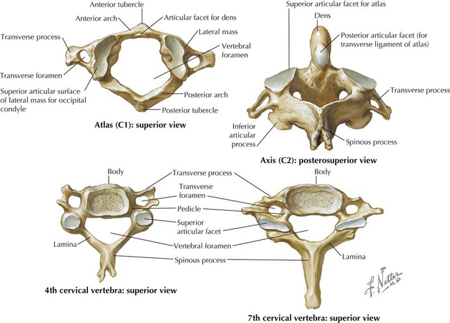
Cervical Vertebrae
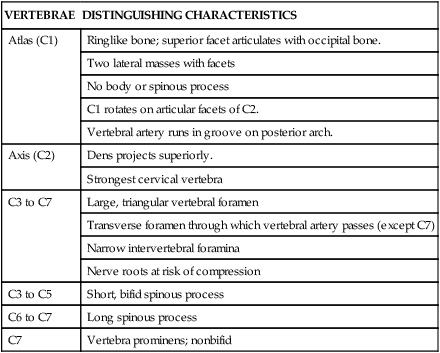
VERTEBRAE
DISTINGUISHING CHARACTERISTICS
Atlas (C1)
Ringlike bone; superior facet articulates with occipital bone.
Two lateral masses with facets
No body or spinous process
C1 rotates on articular facets of C2.
Vertebral artery runs in groove on posterior arch.
Axis (C2)
Dens projects superiorly.
Strongest cervical vertebra
C3 to C7
Large, triangular vertebral foramen
Transverse foramen through which vertebral artery passes (except C7)
Narrow intervertebral foramina
Nerve roots at risk of compression
C3 to C5
Short, bifid spinous process
C6 to C7
Long spinous process
C7
Vertebra prominens; nonbifid
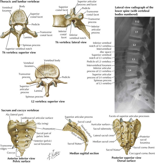
Thoracic and Lumbar Vertebrae
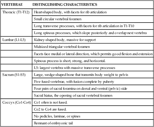
VERTEBRAE
DISTINGUISHING CHARACTERISTICS
Thoracic (T1-T12)
Heart-shaped body, with facets for rib articulation
Small circular vertebral foramen
Long transverse processes, with facets for rib articulation in T1-T10
Long spinous processes, which slope posteriorly and overlap next vertebra
Lumbar (L1-L5)
Kidney-shaped body, massive for support
Midsized triangular vertebral foramen
Facets face medial or lateral direction, which permits good flexion and extension
Spinous process is short, strong, and horizontal.
L5: largest vertebra with massive transverse processes
Sacrum (S1-S5)
Large, wedge-shaped bone that transmits body weight to pelvis
Five fused vertebrae, with fusion complete by puberty
Four pairs of sacral foramina on dorsal and ventral (pelvic) side
Sacral hiatus, the opening of sacral vertebral foramen
Coccyx (Co1-Co4)
Co1 often is not fused.
Co2 to Co4 are fused.
No pedicles, laminae, or spines
Remnant of embryonic tail
Sacrum and Coccyx
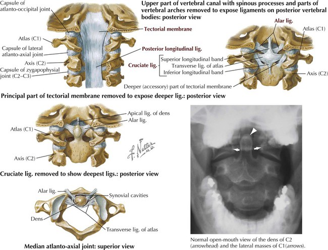
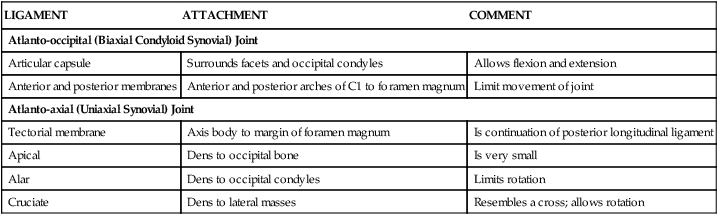
Joints and Ligaments of Craniovertebral Spine
LIGAMENT
ATTACHMENT
COMMENT
Atlanto-occipital (Biaxial Condyloid Synovial) Joint
Articular capsule
Surrounds facets and occipital condyles
Allows flexion and extension
Anterior and posterior membranes
Anterior and posterior arches of C1 to foramen magnum
Limit movement of joint
Atlanto-axial (Uniaxial Synovial) Joint
Tectorial membrane
Axis body to margin of foramen magnum
Is continuation of posterior longitudinal ligament
Apical
Dens to occipital bone
Is very small
Alar
Dens to occipital condyles
Limits rotation
Cruciate
Dens to lateral masses
Resembles a cross; allows rotation
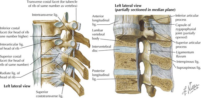
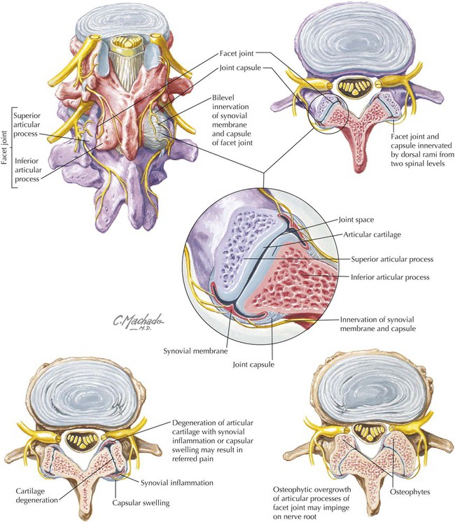
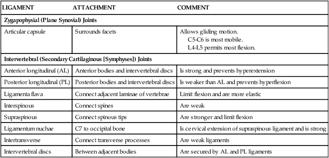
Joints and Ligaments of Vertebral Arches and Bodies
LIGAMENT
ATTACHMENT
COMMENT
Zygapophysial (Plane Synovial) Joints
Articular capsule
Surrounds facets
Allows gliding motion.
C5-C6 is most mobile.
L4-L5 permits most flexion.
Intervertebral (Secondary Cartilaginous [Symphyses]) Joints
Anterior longitudinal (AL)
Anterior bodies and intervertebral discs
Is strong and prevents hyperextension
Posterior longitudinal (PL)
Posterior bodies and intervertebral discs
Is weaker than AL and prevents hyperflexion
Ligamenta flava
Connect adjacent laminae of vertebrae
Limit flexion and are more elastic
Interspinous
Connect spines
Are weak
Supraspinous
Connect spinous tips
Are stronger and limit flexion
Ligamentum nuchae
C7 to occipital bone
Is cervical extension of supraspinous ligament and is strong
Intertransverse
Connect transverse processes
Are weak ligaments
Intervertebral discs
Between adjacent bodies
Are secured by AL and PL ligaments
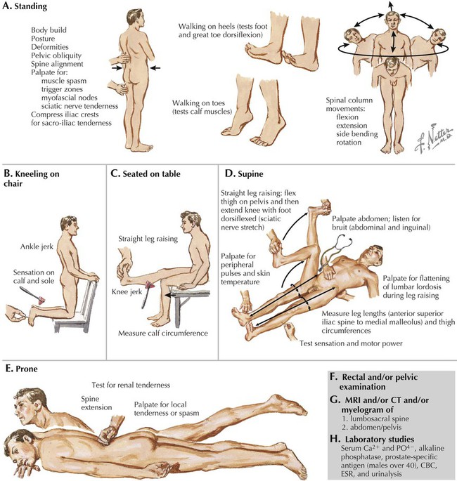
Movements of the Spine
Back