5 Autonomic nervous system
The autonomic nervous system is divided into parasympathetic and sympathetic divisions and these control involuntary activities (see p. 8 and Figs 1.8 and 1.9). Characteristically, autonomic nerves consist of two-neuron chains, with the cell body of the first (preganglionic) lying in the CNS and that of the second (postganglionic) in a ganglion outside the CNS.
Parasympathetic nervous system
The ganglia of the parasympathetic nerves reside within body organs. Preganglionic nerves are generally long while the postganglionic nerves are short, with few interconnections between ganglions and this results in discrete activation of effector cells (see Fig. 1.7).
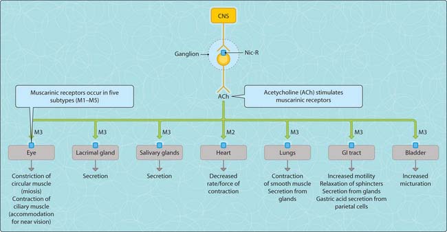
Activation of the parasympathetic nervous system triggers a cascade of events that ends with a biological response (Fig. 3.5.1). The release of acetylcholine from preganglionic neurons activates nicotinic receptors on cell bodies of postganglionic neurons within the ganglia. This activation leads to Na+ entry, membrane depolarization and activation of voltage-dependent sodium channels and it triggers a wave of depolarization along the length of the nerve, ultimately leading to Ca2+ entry at the terminal endings via voltage-dependent calcium channels, fusion of acetylcholine vesicles with the plasma membrane and exocytosis of acetylcholine molecules into the neuroeffector junction. Acetylcholine rapidly activates muscarinic receptors on postjunctional membranes of effector cells to evoke a biological response. The release of acetylcholine from these nerve terminals is controlled by prejunctional muscarinic receptors, which limit further release of acetylcholine (negative feedback mechanism). The pharmacological action of acetylcholine is rapidly terminated by the enzyme acetylcholinesterase, which is expressed on the surface of nerve terminals and postsynaptic membranes.
< div class='tao-gold-member'>
Stay updated, free articles. Join our Telegram channel

Full access? Get Clinical Tree


