Atypical Lipomatous Tumor/Well-Differentiated Liposarcoma
Thomas Mentzel, MD
Key Facts
Terminology
Intermediate (locally aggressive, nonmetastasizing) lipogenic neoplasm composed of atypical adipocytes
Clinical Issues
Accounts for 40-45% of all liposarcomas
Occurs most frequently in deep soft tissues of limbs followed by retroperitoneum, abdominal cavity, paratesticular region, and mediastinum
Usually presents as deep-seated, painless, and slowly enlarging tumor mass
Lesions located in surgically amenable soft tissues recur only rarely after complete excision
Neoplasms arising intraabdominal, retroperitoneum, mediastinum, or spermatic cord often recur repeatedly and may cause death
Variable risk of dedifferentiation in extremities (< 2%) and retroperitoneum (> 20%)
May also arise in subcutaneous tissue and very rarely in skin
Middle-aged to elderly adults
Intermediate (locally aggressive but nonmetastasizing) malignant mesenchymal tumor
Microscopic Pathology
Atypical adipocytes, atypical stromal cells, lipoblasts
Adipocytes show striking variations in size and shape
Enlarged hyperchromatic nuclei
Lipoma-like subtype
Sclerosing subtype
Inflammatory subtype
Spindle cell subtype
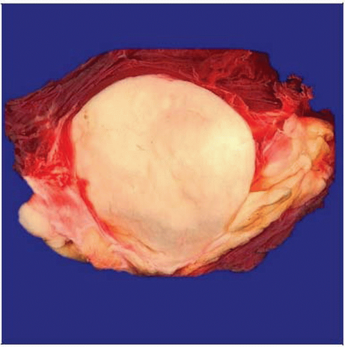 Gross pathology photograph shows a well-circumscribed neoplasm of deep soft tissues with indurated, yellow-white cut surfaces. |
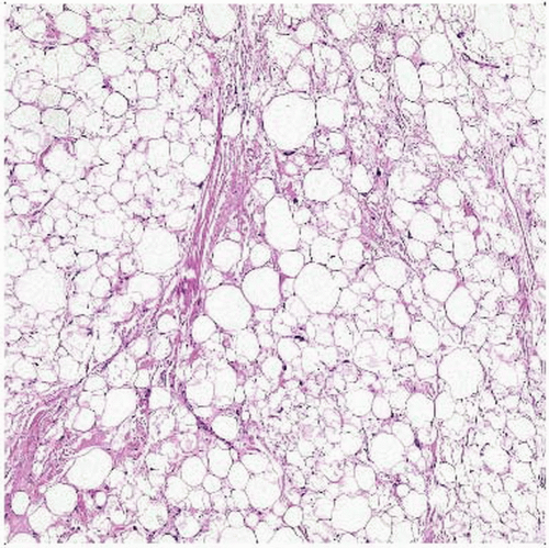 Hematoxylin & eosin shows striking variations in size and shape of lipogenic cells, as well as scattered enlarged cells with enlarged hyperchromatic nuclei. |
TERMINOLOGY
Abbreviations
Atypical lipomatous tumor (ALT)
Synonyms
Well-differentiated liposarcoma (WDLS)
Definitions
Intermediate (locally aggressive, nonmetastasizing) lipogenic neoplasm composed of atypical adipocytes
CLINICAL ISSUES
Epidemiology
Incidence
Accounts for 40-45% of all liposarcomas
Most frequently in deep soft tissues
Retroperitoneum, abdominal cavity, paratesticular region, mediastinum
Limbs
May also arise in subcutaneous tissue and very rarely in skin
Age
Middle-aged to elderly adults
Extremely rare in childhood
Gender
M = F
Presentation
Deep-seated, painless, and slowly enlarging tumor mass
Treatment
Complete surgical excision
Prognosis
In surgically amenable site
Recur only rarely after complete excision
Intraabdominal, retroperitoneal, mediastinal, or paratesticular lesions
Often recur locally and may be fatal
Variable risk of dedifferentiation in extremities (< 2%) and in retroperitoneum (> 20%)
IMAGE FINDINGS
General Features
Best diagnostic clue
Circumscribed lobular mass
Location
Deep soft tissues
Size
Variable
Usually > 5 cm
Morphology
Circumscribed lipomatous lesion
MACROSCOPIC FEATURES
General Features
Well-circumscribed lobular neoplasms
Color varies from yellow to white
Fat necrosis may be seen in large lesions
Sections to Be Submitted
Sample margins and representative sections of tumor
Look for indurated, firm areas
Size
May attain very large size
MICROSCOPIC PATHOLOGY
Histologic Features
Lipoma-like subtype
Adipocytes show significant variation in size and shape
Enlarged hyperchromatic nuclei
Hyperchromatic and multinucleated stromal cells
Lipoblasts may be seen but are not essential for diagnosis
Involvement of large vessel walls by atypical tumor cells
Prominent myxoid stromal changes may be present
Rare chondroid stromal changes
Sclerosing subtype
Scattered bizarre stromal cells with hyperchromatic nuclei
Rare atypical lipogenic cells and multivacuolated lipoblasts
Fibrillary, collagenous stroma
Inflammatory subtype
Prominent inflammatory infiltrate (lymphocytes, plasma cells)
Scattered atypical lipogenic cells/lipoblasts
Often edematous stroma
Spindle cell subtype
Atypical lipogenic cells
Slightly atypical neuroid spindle cells
Fibrous, fibromyxoid stroma
Heterologous differentiation rarely seen
Smooth or striated muscle
Cartilage, bone
Predominant Pattern/Injury Type
Circumscribed
Predominant Cell/Compartment Type
Adipose
Atypical adipocytes, atypical stromal cells, lipoblasts
Grade
Intermediate (locally aggressive but nonmetastasizing) malignant mesenchymal tumor


