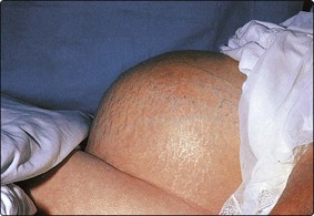
Ascites and pleural fluid
Ascites
Ascites is fluid in the peritoneal space. It can usually be detected by clinical examination (Fig 64.1). Laboratory analysis of ascitic fluid may provide answers to important clinical questions, as its composition varies depending on the underlying cause.
Transudate or exudate?
Traditionally, ascites has been classified based on the protein concentration of the accumulated fluid. Transudates have less protein than exudates. Most cut-offs lie between 20 and 30 g/L. Inflammatory causes of ascites, e.g. malignancy or infection, are usually associated with exudates, whilst transudates more commonly reflect reduced plasma oncotic pressure or increased plasma hydrostatic pressure. However, total protein concentration is not always a reliable guide and comparison of serum and ascites albumin may provide better diagnostic information.
Serum-ascites albumin gradient
The serum-ascites albumin gradient (SAAG) is defined as the serum albumin concentration minus the ascitic fluid albumin concentration. The SAAG correlates directly with the portal pressure. Patients with a wide SAAG (defined as ≥11 g/L) have portal hypertension, whereas patients with a narrow SAAG (<11 g/L) do not (Table 64.1).
Table 64.1
Serum-ascites albumin gradient
Wide (>11 g/L) | Narrow (<11 g/L) |
Chronic liver disease (cirrhosis) | Peritoneal carcinomatosis |
Veno-occlusive disease | Reduced plasma oncotic pressure (e.g. nephrotic syndrome) |
Massive hepatic metastases | Secondary peritonitis |
Congestive cardiac failure | Tuberculous peritonitis |
Spontaneous bacterial peritonitis |
Sometimes causes of ascites that are normally associated with a narrow gradient will occur in patients with portal hypertension, in which case the gradient will be wide. In these situations additional analyses may assist with the differential diagnosis. For example, abnormalities of pH, lactate, glucose and/or lactate dehydrogenase (LDH) may point towards an inflammatory process; increased lymphocytes in the ascitic fluid may point towards tuberculosis, lymphomas or fungal infections of the peritoneum; and malignant cells are found in nearly all patients with peritoneal carcinomatosis (where the tumour directly involves the peritoneum).
Peritonitis
Cirrhotic patients with ascites are prone to develop peritonitis, usually without an obvious focus of infection (so-called spontaneous bacterial peritonitis or SBP). Less commonly, identifiable sources of infection, e.g. perforated viscus or intra-abdominal abscess, are responsible (secondary infection). Laboratory investigations can assist in three ways. First, they may be used to predict who is going to develop SBP. Second, they may permit rapid detection of infection. Third, they may help to differentiate SBP from secondary infection.
Prediction
A low protein concentration in ascitic fluid predisposes to SBP; the ability of macrophages to consume bacteria disappears below a total protein concentration of 10 g/L. In addition, a high bilirubin or a low platelet count identifies individuals who are at particularly high risk.

Full access? Get Clinical Tree

