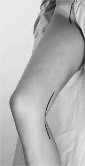
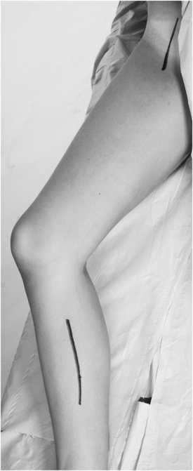
What are the anatomical landmarks of the peripheral pulses in the lower limbs?
Femoral pulse: just inferior to the midinguinal point (halfway between anterior superior iliac spine and pubic symphysis).
Popliteal pulse: bimanual examination; knee slightly flexed, thumbs on tibial tuberosity anteriorly; index fingers palpate pulse deep in the popliteal fossa.
Dorsalis pedis pulse: between first and second metatarsal bones, lateral to the tendon of the extensor hallucis longus.
How are pulses graded?
- 3+
aneurysmal
- 2+
normal
- 1+
present but reduced
- 0
absent
How are lower limb pulses noted?
The example in the diagram shows a patient with an abdominal aortic aneurysm who has sustained an embolic event in his right SFA leading to loss of pulses in the right lower limb below the common femoral artery. Note that he has a full complement of pulses in his left lower limb, which may suggest this is an acute embolic event in the light of the fact that there is no evidence of atherosclerotic disease in the contralateral (left) limb.

What is the most proximal pulse that should be palpated in the arterial examination of the lower limbs?
The aortic pulsation (not the femoral pulse).
How can the diseased arterial segment be deduced by the bypass graft identified?
Axillofemoral or bifemoral bypass: aortic or bilateral common iliac disease.
Femoro-femoral crossover bypass: iliac disease; aorto-iliac aortic endoprosthesis in EVAR.
Femoro-popliteal (above or below knee): superficial femoral or popliteal arterial disease.
Femoro-distal: SFA, popliteal and/or crural arterial disease.

Lower limb arterial scars: bilateral groin cut-down scars are used to expose the common femoral arteries. These can be vertical or oblique.
Causes:
1. EVAR
2. Bilateral femoral endarterectomy
3. Femoral–femoral crossover graft

Lower limb revascularisation scars: aorto-bifemoral bypass.
Can bypass grafts be palpated?
Grafts tunnelled subcutaneously may be palpable. Grafts tunnelled deeply may not be palpable, but the presence of pulses distal to them may suggest that they are patent. Bedside Doppler examination should be used to determine the patency of any graft.
Stay updated, free articles. Join our Telegram channel

Full access? Get Clinical Tree


