CHAPTER 61 Anterior abdominal wall
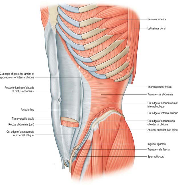
The anterior abdominal wall extends from the costal margins and xiphoid process superiorly to the iliac crests, pubis and pubic symphysis inferiorly. It overlaps and is connected to both the posterior abdominal wall and paravertebral tissues. It forms a continuous but flexible sheet of tissue across the anterior and lateral aspects of the abdomen. The anterior abdominal wall is composed of the integument, muscles and connective tissue lining the peritoneal cavity (Figs 61.1–61.5). It has an important role in maintaining the form of the abdomen and is involved in many physiological activities. Anterior abdominal wall tissues form the inguinal canal that connects the abdominal cavity to the scrotum in men or labia majora in women, and also form the umbilicus; both of these sites are of considerable clinical importance.

Fig. 61.2 Muscles of the left side of the trunk. External oblique has been removed to show internal oblique, but its digitations from the ribs have been preserved. The sheath of rectus abdominis has been opened and its anterior lamina removed.

Fig. 61.3 The cutaneous branches of the lower intercostal and lumbar nerves. Portions of the muscles of the anterior abdominal wall have been removed, including most of the anterior layer of the rectus sheath and parts of rectus abdominis.

Fig. 61.4 Deep muscles and arterial supply of the anterolateral abdominal wall. The greater part of the left rectus abdominis has been removed to show the superior and inferior epigastric vessels.

Fig. 61.5 Left transversus abdominis. The aponeurosis of transversus abdominis fuses into the posterior layer of the rectus sheath above the arcuate line. The position of lateral border of rectus abdominis is shown by the white dotted line.
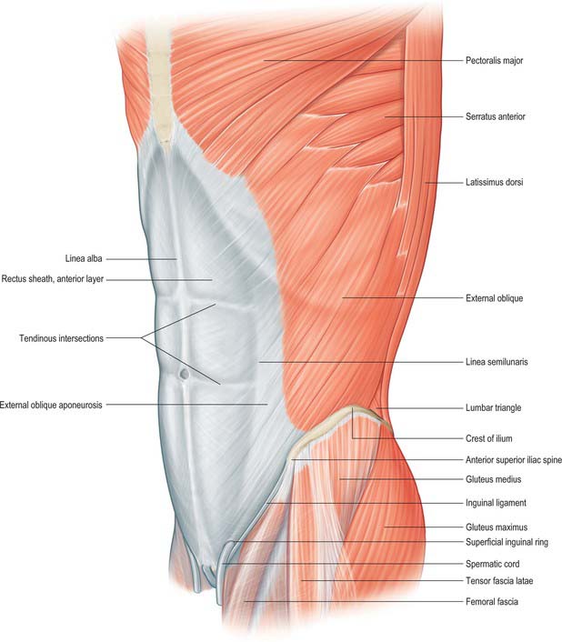
SKIN AND SOFT TISSUE
The integument of the anterior abdominal wall comprises skin, soft tissues, lymphatic and vascular structures, and segmental nerves. The outer layer is formed from the skin and subcutaneous fat. The skin is non-specialized and variably hirsute, depending on the sex and race. All post-pubertal individuals have some extension of the pubic hair onto the anterior abdominal wall skin, although this is commonly most pronounced in males, in whom the hair may extend almost up to the umbilicus in a triangular pattern. The subcutaneous fat of the abdominal wall is highly variable in thickness and is one of the areas where excess fat is stored during periods of obesity, particularly in males.
VASCULAR SUPPLY AND LYMPHATIC DRAINAGE
The anterior abdominal wall receives its blood supply from paired superior and inferior epigastric arteries running vertically through the tissues, and from paired posterior intercostal, subcostal and lumbar vessels running obliquely around the anterolateral aspects of the abdomen.
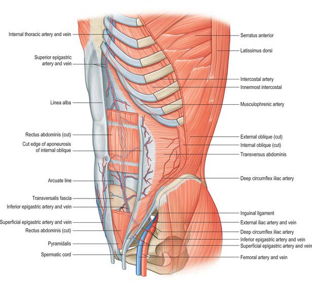
Superior epigastric artery and veins
The superior epigastric artery is a terminal branch of the internal thoracic artery. It descends between the costal and xiphoid slips of the diaphragm, accompanied by two or more veins (Fig. 61.4). The vessels pass anterior to the lower fibres of transversus thoracis and the upper fibres of transversus abdominis. The artery enters the rectus sheath behind rectus abdominis and runs down to anastomose with the inferior epigastric artery usually above the level of the umbilicus. Branches supply rectus abdominis and perforate the sheath to supply the abdominal skin. A branch given off in the upper rectus sheath passes anterior to the xiphoid process of the sternum and anastomoses with the same contralateral branch. This vessel may give rise to troublesome bleeding during surgical incisions that extend up to and alongside the xiphoid process. The superior epigastric artery supplies small branches to the anterior part of the diaphragm. On the right, small branches reach the falciform ligament, where they anastomose with branches arising from the hepatic artery.
Inferior epigastric artery and veins
The inferior epigastric artery originates from the external iliac artery posterior to the inguinal ligament (Fig. 61.6). Its accompanying veins, usually two, drain into the external iliac vein. It curves forwards in the anterior extraperitoneal tissue and ascends obliquely along the medial margin of the deep inguinal ring. It lies posterior to the spermatic cord, but is separated from it by the transversalis fascia. It pierces the transversalis fascia which forms the flimsy posterior support of rectus abdominis, and ascends between the muscle and the fascia and overlying pre-peritoneal connective tissue. In this part of its course, it raises the parietal peritoneum of the anterior abdominal wall as the lateral umbilical fold but has little supporting tissue posteriorly. Disruption of the artery, by surgical incisions e.g. for laparoscopic ports or drains, is not uncommon and the resulting haematoma may expand to considerable size because of the lack of tissue against which the bleeding is effectively compressed. The artery divides into numerous branches. Those which anastomose with branches of the superior epigastric artery do so posterior to rectus abdominis at a variable height above the umbilicus. The inferior epigastric vessels are usually significantly larger than the superior vessels and provide the ‘dominant’ supply to rectus abdominis. Preparatory ligation of the inferior epigastric artery is often performed for myo(cutaneous) flaps using the mid or lower rectus abdominis based on the superior epigastric artery to allow expansion of the superior arterial flow to improve viability of the flap. Branches anastomose with terminal branches of the lower six posterior intercostal arteries posterior to rectus abdominis at the lateral border close to the sheath. The artery is an important inferomedial relation of the deep inguinal ring, and may be damaged during extensive medial dissection of the deep ring during hernia repair, particularly when this is performed in the preperitoneal plane. The vas deferens in the male, or round ligament in the female, wind laterally round it. It has the following branches: the cremasteric artery, a pubic branch, and muscular and cutaneous branches.

Fig. 61.6 A, The deep aspect of the lower part of the abdominal wall of the left side with the thinner posterior wall of the rectus sheath. The femoral and deep inguinal rings are displayed, together with the vessels and other structures in relation to them and also the opening into the obturator canal. B, Laparoscopic view showing the parietal peritoneum covering the area. I, inferior epigastric vessels; H, hernia orifice; D, ductus deferens; T, testicular vessels; E, external iliac vessels.
(B From Drake, Vogl and Mitchell 2005.)
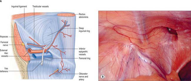
The cremasteric artery accompanies the spermatic cord in males and supplies the cremaster and other coverings of the cord. It anastomoses with the testicular artery. In females it is small and accompanies the round ligament. A pubic branch, near the femoral ring, descends posterior to the pubis and anastomoses with the pubic branch of the obturator artery. Occasionally, the pubic branch of the inferior epigastric artery is larger than the main obturator artery origin, and it supplies the majority of flow into the obturator artery in the thigh. It is then referred to as the aberrant obturator artery. It lies close to the medial border of the femoral ring and may be damaged in medial dissection of the ring during femoral hernia repair. Muscular branches supply the abdominal muscles and peritoneum, and anastomose with the circumflex iliac and lumbar arteries. Cutaneous branches perforate the aponeurosis of external oblique, supply the skin and anastomose with branches of the superficial epigastric artery.
Sometimes the inferior epigastric artery arises from the femoral artery. It then ascends anterior to the femoral vein into the abdomen to follow its course as above. It occasionally arises from the external iliac artery, in common with an aberrant obturator artery and, rarely, from the obturator artery.
The superior and inferior epigastric arteries are important sources for a potential collateral circulation between the internal thoracic artery and the external iliac artery in situations where flow in the thoracic or abdominal aorta is compromised. Small tributaries of the inferior epigastric vein drain the skin around the umbilicus and anastomose with the terminal branches of the umbilical vein, draining the inner surface of the umbilicus via the falciform ligament. These anastomoses may open widely in cases of portal hypertension, when portal venous blood may drain into the systemic circulation via the inferior epigastric vessels. The radiating dilated veins seen under the umbilical skin are referred to as the ‘caput medusae’.
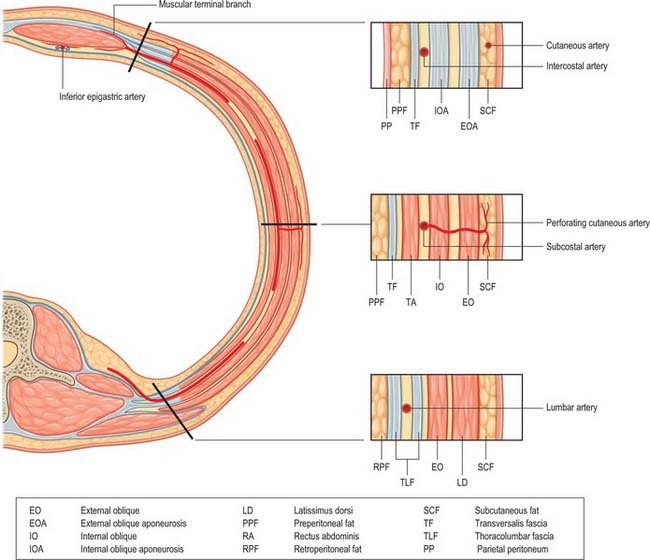
Posterior intercostal, subcostal and lumbar arteries
The 10th and 11th posterior intercostal arteries and the subcostal artery emerge from under the subcostal groove of their respective ribs and pass into the tissues of the anterior abdominal wall. They run through the aponeurosis of transversus abdominis and lie deep to the fibres of internal oblique. The lumbar arteries also cross the aponeurosis of transversus abdominis and lie deep to internal oblique. The arteries on either side run forward, giving off muscular branches to the overlying internal and external oblique, before anastomosing with the lateral branches of the superior and inferior epigastric arteries at the lateral border of the rectus sheath (Fig. 61.7). Perforating cutaneous vessels run vertically through the muscles to supply the overlying skin and subcutaneous tissue. A small contribution to the supply of the lower abdominal muscles comes from branches of the deep circumflex iliac arteries.
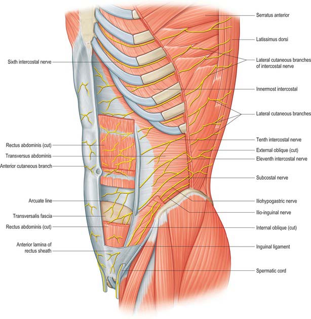
Lymphatic drainage
The lymphatic vessels of the anterior abdominal wall lie both superficial and deep to the deep fascia.
Superficial vessels
The superficial lymphatic vessels accompany the subcutaneous blood vessels. Vessels from the lumbar and gluteal regions run with the superficial circumflex iliac vessels. Those from the infra-umbilical skin run with the superficial epigastric vessels. Both drain into the superficial inguinal nodes. The supra-umbilical region is drained by vessels running obliquely up to the pectoral and subscapular axillary nodes, and there is some drainage to the parasternal nodes.
Deep vessels
The deep lymphatic vessels accompany the deep arteries. The vessels from the posterior portion of the abdominal wall pass with the lumbar arteries to drain into the lateral aortic and retro-aortic nodes. Vessels from the upper anterior abdominal wall run with the superior epigastric vessels to the parasternal nodes. Vessels of the lower abdominal wall drain into the circumflex iliac, inferior epigastric and external iliac nodes.
SEGMENTAL NERVES
The seventh to the 12th lower thoracic ventral rami continue anteriorly from the intercostal spaces into the abdominal wall (Fig. 61.3). Approaching the anterior ends of their respective spaces, the seventh and eighth nerves curve superomedially across the deep surface of the costal cartilages between the digitations of transverse abdominis. They reach the deep aspect of the posterior layer of the aponeurosis of internal oblique. Both the seventh and eighth nerves then run through this aponeurosis, pass posterior to rectus abdominis and supply branches to the upper portion of the muscle. They pass through the muscle near its lateral edge and pierce the anterior rectus sheath to supply the skin of the epigastrium.
The ninth to 11th intercostal nerves pass from their intercostal spaces between digitations of the diaphragm and transversus abdominis. They enter the layer between transversus abdominis and internal oblique. Here, the ninth nerve runs forwards almost horizontally, whereas the tenth and 11th pass inferomedially. At the lateral edge of rectus abdominis, the nerves pierce the posterior layer of the aponeurosis of internal oblique and pass behind the muscle to end, like the seventh and eighth intercostal nerves, with cutaneous branches. The ninth nerve supplies skin above the umbilicus, the tenth supplies skin, which includes the umbilicus, and the 11th supplies skin below the umbilicus (see Fig. 15.12 and Chapters 42, 45 and 79). The 12th thoracic nerve (subcostal nerve) connects with the first lumbar ventral ramus (dorsolumbar nerve). It accompanies the subcostal vessels along the inferior border of the 12th rib, passing behind the lateral arcuate ligament and kidney and anterior to the upper part of the quadratus lumborum. It perforates the transversus abdominis fascia, running deep to the internal oblique, to be distributed like the lower intercostal nerves. It supplies the anterior gluteal skin reaching down to the greater trochanter.
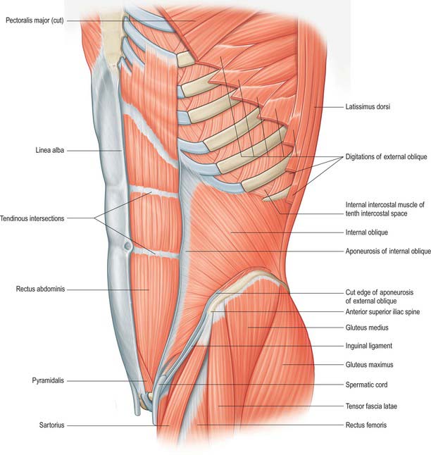
SOFT TISSUE
Superficial fascia
The superficial fascia of the abdominal wall consists mostly of a single layer that contains a variable amount of fat. It lies between the skin and muscles of the anterior abdominal wall. In the lower part, the fascia differentiates into superficial and deep layers between which lie superficial vessels and nerves and, in the region of the groin, superficial inguinal lymph nodes.
Superficial layer
Where present, the superficial layer is thick, areolar in texture and contains a variable amount of fat. It is often greatly thickened in obese individuals. Inferiorly, it lies superficial to the inguinal ligament and is continuous with the superficial fascia of the thigh. In the male, this layer continues over the penis and outer surface of the spermatic cord into the scrotum, where it changes its character. In this region it is generally thin, with very little adipose tissue present and is pale red in colour. In the scrotum it also contains smooth muscle fibres, which form the dartos muscle. From the scrotum it may be traced backwards into continuity with the superficial fascia of the perineum. In the female, it continues from the suprapubic skin of the abdomen into the labia majora and perineum.
Deep (membranous) layer
When present as a distinct entity, the deep layer of the fascia is more membranous than the superficial, and contains elastic fibres. It is loosely connected by areolar tissue to the aponeurosis of external oblique, but in the midline it is intimately adherent to the linea alba and symphysis pubis. In the male, it extends onto the dorsum of the penis and forms part of the superficial ligament of the penis. Superiorly, it is continuous with the superficial fascia over the remainder of the trunk. Inferiorly, it lies superficial to the inguinal ligament and fuses with the overlying superficial layer and the underlying fascia lata in the inguinal flexure line or skin crease of the thigh.
In the male, the deep layer of the fascia continues inferiorly and medially over the penis and spermatic cord to the scrotum, where it becomes continuous with the membranous layer of the superficial fascia of the perineum. In the female, it continues into the labia majora and is continuous with the fascia of the perineum.
In the child, the testis can frequently be retracted out of the scrotum into the loose areolar tissue between external oblique and the deep layer of superficial fascia over the inguinal canal. This ‘space’ is sometimes called the superficial inguinal pouch.
Transversalis fascia
The transversalis fascia is a thin layer of connective tissue lying between the inner surface of transversus abdominis and the extraperitoneal fat. It is part of the general layer of fascia between the peritoneum and the abdominal wall. Posteriorly, it is continuous with the anterior layer of the thoracolumbar fascia, and it forms a continuous sheet anteriorly. Inferiorly, it is continuous with the iliac and pelvic fasciae, and superiorly it blends with the fascial covering of the inferior surface of the diaphragm. It is attached to the entire length of the iliac crest between the origins of transversus abdominis and iliacus and to the posterior margin of the inguinal ligament between the anterior superior iliac spine and the femoral vessels. In the inguinal region it is thick and dense, and augmented by the aponeurosis of transversus abdominis. Medial to the femoral vessels it is thin and fused to the pubis behind the conjoint tendon. Anterior to the femoral vessels, fascia extends down from the fascia transversalis to form the anterior part of the femoral sheath. The fascia is strengthened here by fibres, which arch transversely. Some fibres spread laterally towards the anterior superior iliac spine, some fibres run medially behind rectus abdominis, and some descend to the pubis behind the conjoint tendon. These arched fibres constitute the deep crural arch. The curved fibres of the deep crural arch thicken the inferomedial part of the rim of the deep inguinal ring. The spermatic cord in the male, or the round ligament of the uterus in the female, pass through the transversalis fascia at the deep inguinal ring. This opening is not visible during dissection through the skin because the transversalis fascia is prolonged on these structures as the internal spermatic fascia, but it can be seen from within the abdomen once the peritoneum has been stripped off. The internal spermatic fascia surrounds the testis and blends with the areolar tissue on the parietal layer of the tunica vaginalis: it may contain smooth muscle fibres.
Extraperitoneal connective tissue
The extraperitoneal connective tissue is a layer of areolar tissue lying between the peritoneum and the fasciae lining the abdominal and pelvic cavities. The amount of extraperitoneal tissue varies: it is especially abundant on the posterior wall of the abdomen, particularly around the kidneys, where it contains much adipose tissue; variable in thickness on the anterolateral wall, where it is thin in children and normal adults but often thickened in the obese, particularly in males; scanty in the suprapubic region, above the iliac crest and in the pelvis. Extraperitoneal tissue is continuous with the epimysium of the muscles of the abdominal wall.
MUSCLES
ANTEROLATERAL MUSCLES OF THE ABDOMEN
Rectus abdominis, pyramidalis, external oblique, internal oblique and transversus abdominis constitute the anterolateral muscles of the abdomen. They act together to perform a range of functions, some of which involve the generation of a positive pressure within one or more body cavities. Although many of these activities may occur with no ‘forced assistance’, activities such as expiration, defecation and micturition may be aided by the generation of a positive intra-abdominal pressure. Parturition, coughing and vomiting always require such a positive pressure. Under resting conditions, the tone developed within the muscles provides support for the abdominal viscera and retains the normal contour of the abdomen. The consequences of lack of muscular support can be seen in conditions such as ‘prune belly syndrome’, where there is congenital absence of these muscles.

Stay updated, free articles. Join our Telegram channel

Full access? Get Clinical Tree

