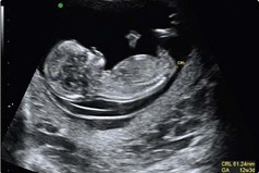
77 (a) Fetal screening for Down’s syndrome and spina bifida. (b) Fetal anomaly screening by ultrasonography – usually at 18–20 weeks – to identify developmental abnormalities, including congenital heart defects and cleft lip and confirm spina bifida. In addition, women are offered screening for HIV, hepatitis B, syphilis and rubella early in pregnancy. Although second trimester screening has been common practice, combined first trimester screening is currently considered to be best practice as it provides a higher detection rate and lower false positive rate. It uses a combination of ultrasound measurement of fetal nuchal translucency (NT), and measurement of the maternal serum markers free beta HCG (FβHCG) and pregnancy-associated plasma protein A (PAPP-A), to derive a combined risk for Down’s syndrome. Each of these markers, including NT, varies with gestation and an accurate measurement of fetal maturity is required for accurate interpretation of results. For first trimester screening, ultrasound measurement of fetal crown rump length (CRL; Fig 77.1), carried out at the same time as the NT measurement, is used as the basis of the calculation of gestation for conversion of marker concentrations into a multiple of the median (MoM). An MoM is a measure of how far an individual test result deviates from the median. MoM is commonly used to report the results of medical screening tests, particularly where the results of the individual tests are highly variable.
Antenatal screening
Overview of screening programmes
First trimester screening
Basicmedical Key
Fastest Basicmedical Insight Engine