Chapter 39 Anoscopy
Common indications
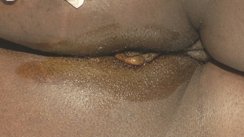
Anoscopy can be used to visualize the anal canal and evaluate anorectal symptoms such as pain or bleeding (Figure 39-1).
Key steps
1. Position patient: Place the patient in a lateral decubitus position with the knees flexed toward the chest. Hold the buttocks apart and visually inspect the external anatomy.
2. Digital examination: After helping the patient relax, lubricate the examining finger and gently perform a digital rectal exam, palpating the anal canal for tone, masses, or tenderness.
3. Insert anoscope: Lubricate the anoscope and the central guide plug, and slowly insert the anoscope through the anus (Figure 39-2).
4. Remove central guide: After the anoscope is completely inserted, remove the central guide, and place it into a container for soiled instruments (Figure 39-3).
5. Inspect mucosa: Slowly rotate the scope as it is withdrawn, and inspect the entire mucosa, looking for mass lesions, hemorrhoids, or fissures (Figure 39-4). Masses or polyps visible through the anoscope may be sampled using a small biopsy forceps.
Stay updated, free articles. Join our Telegram channel

Full access? Get Clinical Tree


