Analytical techniques

Chapter 5 Analytical techniques


It is important that any oil used in aromatherapy is of the highest quality and purity. Essential oils have widespread applications including perfumery, pharmaceutical products and food flavouring. It is estimated that only 5% of all essential oils produced are used in aromatherapy.


As previously stated, aromatherapy-quality oils must be pure and natural, with no manipulation of their composition after extraction. In conventional terms, a chemically pure substance is made up of chemically identical atoms or molecules that have a uniform structure. As essential oils are made up of a mixture of many organic compounds, subject to variation produced by factors such as the growth conditions of the original plants and the extraction processes used, there will be some variation in the composition of oils with the same name.


When analyzing oils, there are a number of techniques that are consistently used. They can give us two types of information:


The main technique used is gas–liquid chromatography (abbreviated GLC or nowadays just GC), which is especially useful when combined with mass spectroscopy (MS); the combination is often referred to as GC-MS. They can provide both quantitative and qualitative information that is very accurate and reliable when compared to known analytical measurements of oils that are stored in databases.


CHROMATOGRAPHY


Chromatography refers to a range of closely related techniques used for separating mixtures. Chromatography is used extensively in analysis of mixtures in a wide range of applications. Many different techniques have been developed to identify specific compounds in specialist applications.


All chromatography relies on a mobile phase moving through a stationary phase. The components of the mixture are attracted to both phases and become distributed between them. It is the differing relative strength of attraction of the components for the two phases that is important. If a component is strongly attracted to the stationary phase, it will be held back, while one with a strong attraction for the mobile phase will quickly move along with it. The choice of materials in the stationary and mobile phases must be suitable to allow the components of the mixture to move at different speeds and thus be separated.


The rate at which a component moves will depend on its equilibrium concentrations in mobile and stationary phases. By the technical term equilibrium concentrations we are essentially referring to the solubilities of that component in each of the phases. The ratio of these concentrations is known as the distribution coefficient (symbolized D).


image


The substance must be in the same molecular form in both phases for this relationship to apply (i.e. we must be comparing the concentrations of identical forms). This D value will always be the same for a given component distributed between a particular combination of phases. This means that the separation can be reproduced to give the same result with the same substance when using the same system.


Gas–liquid chromatography (GC or GLC)


In GC the mobile phase is a gas and the stationary phase is a liquid. It is one of the most widely used techniques for separation of materials and for analysis and can give both qualitative and quantitative information about a sample.


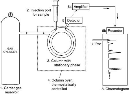
A typical GC apparatus is shown in Figure 5.1.


The gas chromatograph can be calibrated by using a series of known or standard solutions to produce a calibration graph so that the retention times of peaks from the known sample can be compared with the unknown sample. The chromatogram can be examined for the number of peaks produced and peaks can be identified by their retention times. The quantity of compound present in each peak can be found by measuring the area under the peak.


A typical GC analysis chart is shown for a geranium oil in Figure 5.2.


image

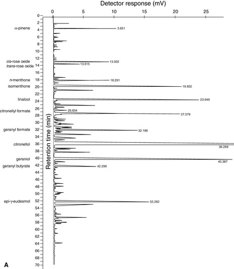
Figure 5.2 Results of GC of geranium oil. (A) This shows the chromatogram, or printed chart, in an analysis that has run for 70 minutes (see the horizontal axis). (B) These are the type of data you would expect to get along with the chart. (Column 1) Peak number in order of retention times – the lower the number, the quicker the compound passes through the column, i.e. the faster it moves in the mobile phase. The order of the peaks represents the volatilities of the compounds: the monoterpenes come off first, sesquiterpenes and their oxygenated compounds in the middle, with the compounds of low volatility last. (Column 2) Peak name identifies the compound. (Column 4) Retention time is the time the vaporized compound takes to pass through the column. (Column 3) Result percentages. These figures are measures of the areas of individual peaks expressed as a percentage of the total area of all of them. This means that all the individual peak areas should add up to 100%. The figures are calculated automatically by a special computer called an integrator. In practice the relative area of each peak is not precisely proportional to the percentage of the corresponding constituent in the essential oil; this has to be worked out using an additional response factor for the substance. For this sample the results show identification of 13 major components out of 289, making up 81.25% of the total components. The volatile monoterpene -pinene (peak 1) is the first off the column. The alcohols citronellol (10) and geraniol (11) are responsible for the odour characteristics of geranium, which is lifted and activated by the two rose oxides (2 and 3). The 6,9-guaiadiene (7) is a non-terpene hydrocarbon that acts as a back note but it is not a powerful odour. Component 12 is geranyl butyrate; component 13 is epi- -eudesmol. Chromatograms and data supplied by Jenny Warden of Traceability.


The gas chromatograph is particularly useful when it is linked to a mass spectrometer. This combination is called gas chromatography–mass spectrometry (GC-MS).


MASS SPECTROMETRY (MS)


Mass spectroscopy is particularly useful for elucidating the components of essential oils. It can determine relative atomic masses, molecular masses (relative molecular weights) and, in the more powerful instruments, obtain molecular formulae with sufficient accuracy for unambiguous identification. Also, each substance produces a fragmentation pattern in the mass spectrometer. This can be used to distinguish between closely related molecules by comparing their fragmentation patterns. These patterns can be used to identify known substances by a ‘fingerprinting’ technique or to give evidence for the arrangement of the atoms in a compound. Thousands of mass spectra are stored in computer databases. These are used so that rapid and accurate comparisons can be made and samples identified.


With the development of high resolution mass spectroscopy the mass of the molecular fragment can be measured to seven significant figures. These very accurate relative atomic masses make it possible to distinguish molecules with very similar molecular mass values.


In a mass spectrometer a compound is vaporized and its molecules are bombarded with high-energy electrons. In collisions the electrons transfer energy to the molecules: the molecules ionize and positive ions are formed (a positive ion is a molecule that has lost one or more electrons, but is otherwise unchanged). The bombarding electrons have enough energy to break the covalent bonds in the molecules, and the molecules or ions fragment into smaller positively charged ions.


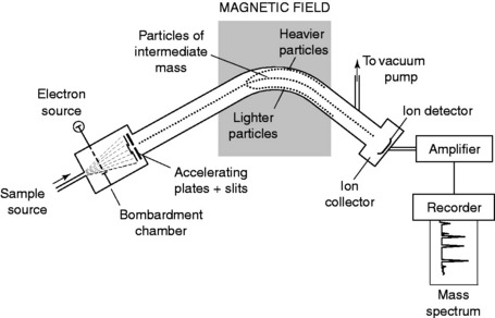
The ions produced are then formed into a beam and accelerated in a magnetic field and deflected by another magnetic field. For ions with the same charge, the deflection in the magnetic field is greatest for ions of lowest mass. The degree of deflection also depends on the amount of charge on the ion – more highly charged ions will be deflected more than ions of the same mass with a lower charge. These two factors are taken into account by the mass spectrometer, which records the relative abundance of each type of particle in terms of its mass (m) to charge (z) ratio (m/z).


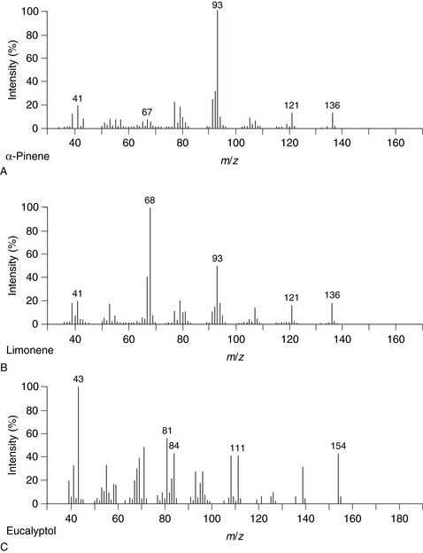
Figure 5.3 shows the typical arrangement of a mass spectrometer. Figure 5.4 shows MS charts for the two monoterpenes [ ]-pinene and limonene, and the oxide 1,8-cineole.


Stay updated, free articles. Join our Telegram channel

Apr 2, 2017 | Posted by in GENERAL SURGERY | Comments Off on Analytical techniques

Full access? Get Clinical Tree

Get Clinical Tree app for offline access