Ampullary Adenoma
Mari Mino-Kenudson, MD
Key Facts
Clinical Issues
Most are sporadic
Seen in 50-95% of patients with (FAP) and Gardner syndrome
Macroscopic Features
May involve one or more parts of ampullary/periampullary region
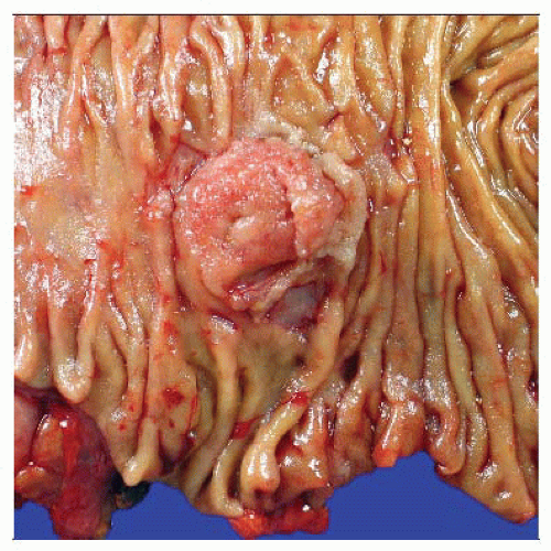
Polypoid, sessile, or subtle mucosal thickening
Microscopic Pathology
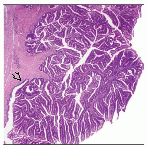
3 histologic categories: Tubular, tubulopapillary (tubulovillous), and papillary (villous)
Papillary almost always associated with invasive adenocarcinoma
Extension of adenomatous epithelium into periductal glands of bile &/or pancreatic ducts or into submucosal glands may mimic invasion
 A single circumscribed mass with a nodular surface occupies the ampulla. The histology of this lesion showed a tubulopapillary adenoma with invasive adenocarcinoma. |
TERMINOLOGY
Definitions
Intestinal-type adenoma of ampullary or periampullary region
Resembles adenomas of small and large bowel
CLINICAL ISSUES
Epidemiology
Incidence
55% of small intestinal adenomas
Most are sporadic
Seen in 50-95% of patients with familial adenomatous polyposis (FAP) and Gardner syndrome
Most common site of extracolonic polyps in FAP patients
Age
Range: 30-80 years
Average: 60s
Polyposis syndrome patients develop them at younger age
Gender
M:F ratio = 1:2.6 in sporadic setting
No gender predominance in polyposis syndromes
Presentation
Symptoms associated with biliary obstruction
Stay updated, free articles. Join our Telegram channel

Full access? Get Clinical Tree


