214 Age-related macular degeneration (senile macular degeneration)
Salient features
History
• Often visual loss is detected when one eye is covered for testing visual acuity
• Loss of ability to read, recognize faces or drive a car; however, patients have enough peripheral vision to walk unaided
• Decrease in visual acuity (severe loss suggests choroidal neovascularization)
• Metamorphosia (distortion of the shape of objects in view)
• Variable visual loss from atrophy of a large area of retinal pigment epithelium involving the fovea
Examination
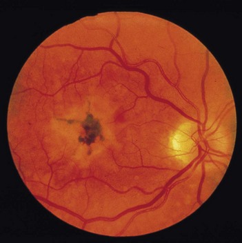
• Disruption of pigment of the retinal pigment epithelium into small areas of hypopigmentation and hyperpigmentation (Fig. 214.1)
• Comment on the white walking aid by the bedside, which indicates the patient is registered blind.
• Check visual acuity and visual field (in most patients there is loss of central vision and maintenance of peripheral vision). Patients with only drüsen typically require additional magnification of text and more intense light to read small print text.
• Tell the examiner you would like to use Amsler grid to confirm your diagnosis.
Questions
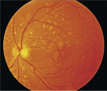
What are drüsen?
Drüsen are pale yellow spots that occur individually or in clusters throughout the macula (Fig. 214.2). Nearly all individuals over the age of 50 years of age have at least one small drüsen (≤63 µm) in one or both eyes (Opthalmology 1992;14:130–42). They consist of amorphous material accumulated between the Bruch’s membrane and pigment epithelium. Although the exact origin is not known, it is believed that drüsen occur from accumulation of lipofuscin and other cellular debris derived from cells of the retinal pigment epithelium that are compromised by age and other factors. Only eyes with large drüsen (>63 µm) are at increased risk for senile macular degeneration (Opthalmology 1997;104:7–21). The clinical hallmark and usually the first clinical finding of age-related macular degeneration is the presence of drüsen. In most cases of age-related macular degeneration, drüsen are present bilaterally.
What are the types of age-related macular degeneration?
There are three types (Lancet 2008; 372:1835–45):
Early age-related macular degeneration. Multiple small drüsen (<63 µm) or intermediate drüsen (63–125 µm) with no evidence of advanced age-related macular degeneration.
Intermediate age-related macular degeneration. Extensive intermediate drüsen or large drüsen (≥125 µm) with no evidence of advanced age-related macular degeneration.
Advanced age-related macular degeneration. The presence of one or other of geographic atrophy or neovascular age-related macular degeneration:
Stay updated, free articles. Join our Telegram channel

Full access? Get Clinical Tree


