Problem 22 Acute back pain in a 75-year-old man
You are asked to see a 75-year-old retired electrician who has been brought into the emergency department with an 18-hour history of increasing back pain. He tells you that the pain came on gradually and now goes down into his left groin and thigh. The pain is constant, getting worse. He has never had this pain before. He does not think the pain was precipitated by anything and nothing seems to ease it. In particular, there is no history of trauma, and walking or exertion do not affect it. There are no other associated symptoms.
Eight months ago he suffered a myocardial infarction and since that time has suffered with moderate angina on effort. He takes isosorbide dinitrate 10 mg three times a day for the angina and uses nitrate skin patches at night. He used to smoke 20 cigarettes a day, but ceased after his heart attack. He drinks 20 g of alcohol per day at the weekends. He suffers occasional episodes of indigestion for which he takes an over-the-counter antacid preparation.
The review of systems is otherwise unremarkable apart from symptoms of increasing bladder neck outflow obstruction, which have been worse over the last 2 years. He gets up twice every night to void and says he feels that he can never empty his bladder properly. He has not passed urine since the pain started.
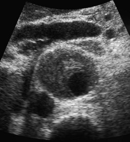
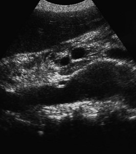
The patient recently had an abdominal ultrasound for his urinary frequency (Figures 22.1 and 22.2).
The ultrasound findings confirm your clinical suspicions.
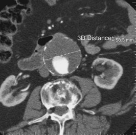
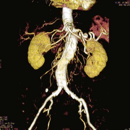
As this patient is haemodynamically stable, a further investigation is performed expeditiously. Two of the images are shown (Figures 22.3 and 22.4).
Q.5
What type of images are these and what do they show? How do these findings affect your management?
Stay updated, free articles. Join our Telegram channel

Full access? Get Clinical Tree


