A middle-aged man with sudden visual loss

Problem 40 A middle-aged man with sudden visual loss


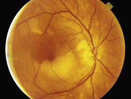
On examination, you measure the blood pressure at 150/95 mmHg. There is no evidence of postural hypotension. Vision, visual fields, pupillary responses, anterior segment examination, gonioscopy and applanation tonometry findings are normal. Visualization of the retina with a direct ophthalmoscope reveals the finding illustrated in Figure 40.1.


After completing the clinical examination, you arrange carotid Doppler ultrasonography. This indicates a severe (85%) stenosis of the origin of the right internal carotid artery. There is also mild stenosis (no more than 50%) in the region of bifurcation of the left common carotid artery.


After evaluating the results of all investigations, you discuss possible medical treatment and surgical intervention with the patient. The patient tells you that on the evening prior to the review appointment, while he was sitting watching television, his left arm became weak and he was unable to pick up his coffee cup. On trying to stand, he found his left leg was dragging, and he was forced to sit down. The episode lasted approximately 10 minutes.


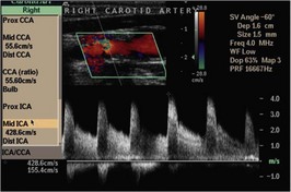
The patient decides in favour of the suggested surgical procedure. In preparation for this, the surgeon orders the radiological study illustrated in Figure 40.2.


The patient undergoes successful carotid surgery. Postoperatively he is maintained on aspirin and is referred to the cardiac rehabilitation service to improve his lifestyle.


Answers


A.1 This patient has presented with transient monocular visual loss (TMVL) or ‘amaurosis fugax’. TMVL is characterized by sudden onset of unilateral visual loss lasting no longer than 24 hours. Commonly of ischemic aetiology, the vascular condition can be in the eye or its vicinity, or can be remote.


TMVL can be due to impaired arterial perfusion (retinal, choroidal or optic nerve ciliary arteries) or to venous stasis. Impending central retinal vein occlusion and central retinal vein occlusion can present with TMVL.


Common causes include arteriosclerosis, hypercoagulable and hyperviscosity states. Though rare, vasculitis and retinal migraine (vasospasm of the retinal arteries) can present with transient visual loss. It is important to consider the possibility of temporal arteritis which may involve the ophthalmic arteries, and has the potential to lead rapidly to arterial occlusion and devastating bilateral blindness. This is most often seen in elderly patients, and other symptoms may include headache, scalp tenderness often apparent when brushing the hair, and jaw claudication, which may occur on chewing food. Polymyalgia rheumatica may co-exist.


TMVL that lasts only for a few seconds is called transient visual obscurations and is typically seen in optic disc oedema (with or without drusen) and in congenital anomalies of the optic disc. The most frequent remote cause for TMVL is carotid stenosis (due to atherosclerosis) rather than cardiac source emboli. The site of carotid stenosis is usually at the termination or at the bifurcation of the common carotid artery or at the origin of the internal carotid artery. Cardiac emboli are associated with atrial fibrillation, mural thrombi (after myocardial infarction) and diseased valves. Other remote causes includes stenosis of the aortic arch and ophthalmic arteries, systemic hypotension and diminished cardiac output. Rarely carotid artery dissection can present with TMVL and may be associated with neck pain and Horner’s syndrome.


Ocular disorders such as intermittent angle closure glaucoma, hyphaema, optic nerve demyelination (Uthoff’s phenomenon) and keratoconus can also mimic transient visual loss and need to be ruled out by ophthalmologic evaluation.


A.2 Figure 40.1 illustrates an embolus at the branch of a retinal artery.


This is most likely a cholesterol embolus or ‘Hollenhorst plaque’, originating from the carotid arterial system.


When evaluating a patient with transient monocular visual loss, a careful cardiovascular examination is essential, including measurement of pulse and blood pressure, examination of carotid and temporal arteries, and assessment of the heart. Remember that the absence of a carotid bruit does not rule out significant carotid disease. Indeed, the most severely diseased carotids often do not have an associated bruit. This patient has an elevated blood pressure, possibly reflecting poor compliance with antihypertensive medication, which is a risk factor for carotid atherosclerosis.


A.3 Essential investigations should include:


Stay updated, free articles. Join our Telegram channel

Apr 2, 2017 | Posted by in GENERAL SURGERY | Comments Off on A middle-aged man with sudden visual loss

Full access? Get Clinical Tree

Get Clinical Tree app for offline access