A 63-year-old woman with a screen-detected abnormality

Problem 9 A 63-year-old woman with a screen-detected abnormality


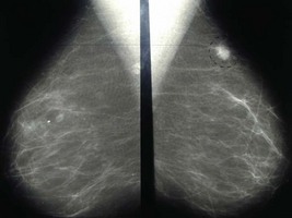
The mammogram is reviewed (Figure 9.1).


An ultrasound examination is performed and shows a hypoechoic lesion with irregular margins, distorting and invading the surrounding breast tissue.


A fine needle aspiration (FNA) is arranged. In the outpatient clinic and with ultrasound guidance a 23 gauge needle is passed several times through the lesion. The aspirate is spread on a microscope slide and examined immediately. Benign cells with atypia are seen, but there is no evidence of malignancy.


A core biopsy is arranged. This procedure requires the use of an automated biopsy gun and 12–18 G biopsy needle. The biopsy is performed under local anaesthesia using ultrasound guidance. A core of architecturally intact breast tissue is removed and invasive ductal carcinoma of intermediate nuclear grade is confirmed.


In this particular case, staging investigations (liver function tests, CT scan chest and abdomen, whole body bone scan) did not reveal any evidence of metastatic disease.


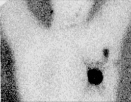
A surgical procedure is planned for the patient. The following investigation is performed immediately before the operation (Figure 9.2).


The patient undergoes wide local excision, specimen X-ray and sentinel node biopsy. Histopathology demonstrates a 22 mm grade II ductal carcinoma with focal vascular invasion. Tumour excision is complete with the closest resection margin being 5 mm from the inferior edge. Of the three sentinel lymph glands removed, two demonstrated metastatic tumour deposits. The patient subsequently underwent completion axillary dissection. Histopathology of the axillary fat demonstrated 1/13 further lymph glands with metastatic tumour. The cancer stained strongly for oestrogen receptor and progesterone receptor but was negative for HER-2 (human epidermal growth factor receptor-2).


The patient underwent chemotherapy followed by sequential breast radiotherapy and hormone treatment using an aromatase inhibitor. Regular follow-up was arranged to include clinical examination and annual mammography.


Answers


A.1 The two most obvious risk factors are that the patient is female and over the age of 50 years. Breast cancer is a disease almost exclusively affecting women although approximately 1% of all breast cancers affect men.


Family history is an important breast cancer risk factor. The nature of the family history is important whereby closely related relatives, bilaterality, age of onset and the number of affected relatives all play a role in determining breast cancer risk. The presence of other organ-specific cancers (i.e. ovarian, soft tissue malignancies) is also important and may indicate a hereditary form of the disease involving breast cancer-specific genes such as BRCA1 or BRCA2.


Hormonal factors also affect breast cancer risk. Women who start menstruating early in life, have a late menopause or have a late age of first pregnancy are at increased risk of developing breast cancer. Current users of hormone replacement therapy (especially combined preparations using both oestrogen and progesterone) are also at increased risk of breast cancer. The risk increases with longer duration of hormone replacement therapy use. The risks associated with HRT disappear 5 years after stopping this treatment.


A.2 The following features on the mammogram would suggest malignancy:


The following features are more suggestive of a lesion being benign:


This woman’s mammogram shows:


These features are suggestive of a malignant lesion and the patient will require further investigtion.


A.3 The woman should be formally assessed by a medical practitioner using the triple test. This includes:


Stay updated, free articles. Join our Telegram channel

Apr 2, 2017 | Posted by in GENERAL SURGERY | Comments Off on A 63-year-old woman with a screen-detected abnormality

Full access? Get Clinical Tree

Get Clinical Tree app for offline access