A 59-year-old man with calf pain

Problem 23 A 59-year-old man with calf pain


Three years previously he suffered a myocardial infarction and since then has been on enteric-coated aspirin. He takes no other medications apart from a salbutamol inhaler which was prescribed by his general practitioner for ‘when he gets a bit tight’. In the past he has had an appendectomy and a prostatectomy. He smokes 20 cigarettes a day and drinks 20 g of alcohol a day. He is 170 cm tall and weighs 85 kg.


His blood pressure is 150/85 mmHg and his heart rate is 84 and regular. Examination of his heart is unremarkable. He has a bruit over his right carotid artery. His chest is resonant with decreased breath sounds and a prolonged expiration, but no added sounds. Abdominal examination is normal. His femoral pulses are palpable, as are the more distal pulses in his left leg. His right popliteal pulse is not palpable and neither are the right pedal pulses. Both legs are of similar temperature and there are no other signs of limb ischaemia.


His ankle–brachial index is 0.5 on the right and 0.8 on the left. His lipid profile is reported as being marginally raised and a random blood sugar is within normal limits. His serum biochemistry is unremarkable.


Twelve months later he comes back to see you. He had managed to continue his daily walks down to the local shops until 2 months ago, but despite your advice he has continued to smoke. His pain has worsened recently and now comes on at about 20 metres. He now finds these symptoms interfere with his lifestyle. Most recently he has been woken at night by pain in his leg, which he relieves by dangling the leg over the edge of the bed.


Your examination of his cardiorespiratory system does not reveal any new findings. His abdomen is soft, there are no pulsatile masses and both femoral pulses are palpable. His legs and feet are cool to the touch, and the right foot is colder than the left. The toes on his right foot are dusky and there is sluggish capillary return to his big toe. When the leg is lowered over the side of the examination couch it assumes a reddish-purple colour, and when the leg is elevated the foot becomes pale. His popliteal and pedal pulses cannot be palpated on either leg. His Doppler pressures are measured and he has an ankle–brachial index of 0.3 on the right and 0.7 on the left.


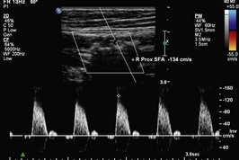
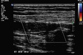
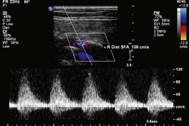
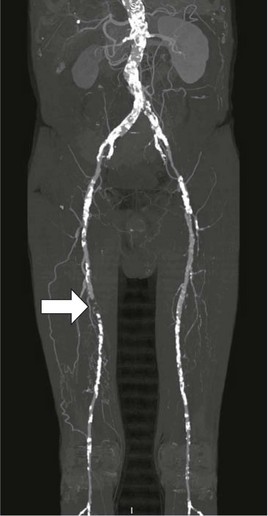
On your advice he agrees to see a vascular surgeon, who recommends that an arterial duplex scan is performed. This is shown in Figures 23.1, 23.2 and 23.3. Figure 23.2 demonstrates an occluded segment of superficial femoral artery (SFA). The waveforms above this lesion (23.1) are normal but the waveforms distally (23.3) are damped consistent with the occlusion just proximal to this segment. Subsequently he recommends a CT angiogram (shown in Figure 23.4). These are AP views with the patient’s right leg on the left side of Figure 23.1.


There is diffuse calcification of all vessels. There is an occlusion of the right distal superficial femoral and proximal popliteal artery confirmed with good below-knee vessels.


An attempted angiography was unsuccessful and a reverse vein femoropopliteal bypass was performed. This is successful in improving his lower limb circulation but is complicated by a cerebrovascular event involving his non-dominant hemisphere for which he requires 3 months of intensive rehabilitation.


Apr 2, 2017 | Posted by in GENERAL SURGERY | Comments Off on A 59-year-old man with calf pain

Full access? Get Clinical Tree

Get Clinical Tree app for offline access