A 41-year-old man involved in a car crash

Problem 26 A 41-year-old man involved in a car crash


Your examination reveals the following:


Airway (with cervical spine control): the patient’s airway is patent, he is speaking to you and has no evidence of facial trauma or airway foreign body. His cervical spine is immobilized with a hard collar.


Breathing: he has a respiratory rate of 30 bpm and his pattern of breathing is shallow. He is not centrally cyanosed. Examination of his chest reveals superficial abrasions and contusion to his left anterior hemithorax and shoulder. There is no evidence of paradoxical chest movement or open wound. He is tender over the anterior aspects of the left 6th–9th ribs and crepitus is present. There is a paucity of breath sounds on the left side and the percussion note is increased on that side. His trachea is central. Pulse oximetry measurement reveals his oxygen saturation is 99% on 15 litres of O2 by non-rebreather mask.


Circulation: he has a pulse rate of 130/min and a blood pressure of 105/85 mmHg. His jugular venous pressure is not elevated and he has a peripheral capillary return time of 3–4 seconds. He has no obvious external haemorrhage or major long bone fractures. The examination of his abdomen reveals bruising consistent with a seatbelt injury and generalized tenderness of both upper quadrants, maximally on the left, with marked guarding.


Disability: a rapid skeletal and neurological survey shows that the patient has no obvious deformity or tenderness of his limbs, has normal power in all limbs and has normal sensation and reflexes. His Glasgow Coma Scale score (GCS) is 14/15, with his verbal score being confused (4/5). His pupils are equal and react normally to light. You also note the odour of alcohol. Breath alcohol analyser shows a breath alcohol level (BAL) of 0.210 g/dL. He has no neck pain or midline cervical tenderness.


Exposure/environmental control: the patient’s temperature is 36.1°C. In order to facilitate examination and X-rays his shirt and trousers’ are cut off. He complains that he is cold in the resuscitation room.


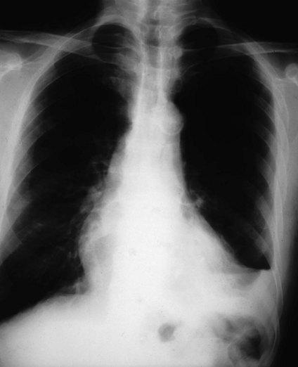
A second wide-bore intravenous cannula is inserted into a peripheral vein and 1 L of a crystalloid solution administered rapidly. Blood samples are collected for cross-matching, complete blood picture and biochemistry. In addition forensic blood alcohol samples are obtained. The chest X-ray is shown in Figure 26.1.


The chest X-ray confirms your clinical suspicion of a haemopneumothorax. A chest drain is inserted successfully and connected to an underwater seal. Air and approximately 500 mL blood drain out. The patient confirms that his breathing is much improved.


A log roll and inspection of the patient’s back is subsequently performed which is unremarkable. FAST scan done on arrival is equivocal.


The initial laboratory results are also unremarkable and his haemoglobin is 131 g/L.


One hour after admission and initial resuscitation you reassess the patient and note that although he has received a total of 2 L intravenous fluid and his pain is adequately controlled with parenteral opiates, his pulse has risen to 145/min and his blood pressure is 90/75 mmHg. His JVP is not visible. His airway and breathing are stable. Examination of his chest shows reasonable air entry to all areas, but there is still dullness at the left base. There is a further 100 mL fresh blood in the chest drainage bottle. His abdomen is markedly tender in the left upper quadrant.


A further haemoglobin estimation is performed. It is now 102 g/L. He is rapidly transfused 2 units blood and a further 1 L crystalloid. His urine dipstick is negative for blood and a urinary catheter is inserted with hourly urine output monitored. His blood pressure stabilizes at 105/80 mmHg with a pulse of 120 bpm.


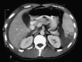
A CT scan of his head, cervical spine, chest, abdomen and pelvis is performed. A slice from the upper abdomen is shown in Figure 26.2.


The patient is transfused a further 2 units of blood. He remains stable, his tachycardia settles and so it is decided to trial treating his splenic injuries conservatively.


He is admitted to an intensive care unit where he is monitored closely and has an uneventful course. His chest drain is removed at day 5. His splenic injury is monitored with a further CT, which showed gradual reabsorption of the haematoma. He is discharged home 2 weeks after the accident and advised to avoid strenuous exertion for a further month.


Apr 2, 2017 | Posted by in GENERAL SURGERY | Comments Off on A 41-year-old man involved in a car crash

Full access? Get Clinical Tree

Get Clinical Tree app for offline access