A


A


Abdominal distention

Abdominal distention refers to increased abdominal girth—the result of increased intraabdominal pressure forcing the abdominal wall outward. Distention may be mild or severe, depending on the amount of pressure. It may be localized or diffuse and may occur gradually or suddenly. Acute abdominal distention may signal life-threatening peritonitis or acute bowel obstruction.

Abdominal distention may result from fat, flatus, a fetus (pregnancy or intra-abdominal mass [ectopic pregnancy]), or fluid. Fluid and gas are normally present in the GI tract but not in the peritoneal cavity. However, if fluid and gas are unable to pass freely through the GI tract, abdominal distention occurs. In the peritoneal cavity, distention may reflect acute bleeding, accumulation of ascitic fluid, or air from perforation of an abdominal organ.

Abdominal distention doesn’t always signal pathology. For example, in anxious patients or those with digestive distress, localized distention in the left upper quadrant can result from aerophagia—the unconscious swallowing of air. Generalized distention can result from ingestion of fruits or vegetables with large quantities of unabsorbable carbohydrates, such as legumes, or from abnormal food fermentation by microbes. Don’t forget to rule out pregnancy in all females with abdominal distention.

image 
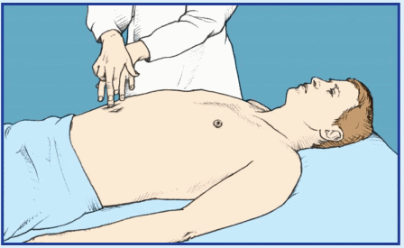
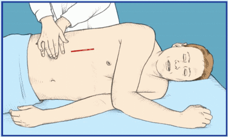
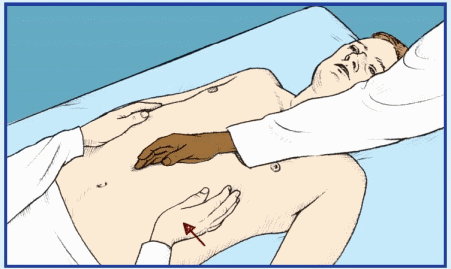
If the patient displays abdominal distention, quickly check for signs of hypovolemia, such as pallor, diaphoresis, hypotension, rapid and thready pulse, rapid and shallow breathing, decreased urine output, poor capillary refill, and altered mentation. Ask the patient if he’s experiencing severe abdominal pain or difficulty breathing. Find out about any recent accidents, and observe the patient for signs of trauma and peritoneal bleeding, such as Cullen’s sign or Turner’s sign. Then auscultate all abdominal quadrants, noting rapid and highpitched, diminished, or absent bowel sounds. (If you don’t hear bowel sounds immediately, listen for at least 5 minutes.) Gently palpate the abdomen for rigidity. Remember that deep or extensive palpation may increase pain.

If you detect abdominal distention and rigidity along with abnormal bowel sounds, and the patient complains of pain, begin emergency interventions. Place the patient in the supine position, administer oxygen, and insert an I.V. catheter for fluid replacement. Prepare to insert a nasogastric tube to relieve acute intraluminal distention. Reassure the patient and prepare him for surgery.


HISTORY AND PHYSICAL EXAMINATION

If the patient’s abdominal distention isn’t acute, ask about its onset and duration and associated signs. A patient with localized distention may report a sensation of pressure, fullness, or tenderness in the affected area. A patient with generalized distention may report a bloated feeling, a pounding heartbeat, and difficulty breathing deeply or breathing when lying flat. The patient may also feel unable to bend at his waist. Be
sure to ask about abdominal pain, fever, nausea, vomiting, anorexia, altered bowel habits, and weight gain or loss.

Obtain a medical history, noting GI or biliary disorders that may cause peritonitis or ascites, such as cirrhosis, hepatitis, or inflammatory bowel disease. (See Detecting ascites.) Also note chronic constipation. Has the patient recently had abdominal surgery, which can lead to abdominal distention? Ask about recent accidents, even minor ones, like falling off a stepladder.

Perform a complete physical examination. Don’t restrict the examination to the abdomen because you could miss important clues to the cause of abdominal distention. Next, stand at the foot of the bed and observe the recumbent patient for abdominal asymmetry to determine if distention is localized or generalized. Then assess abdominal contour by stooping at his side. Inspect for tense, glistening skin and bulging flanks, which may indicate ascites. Observe the umbilicus. An everted umbilicus may indicate ascites or an umbilical hernia. An inverted umbilicus may indicate distention from gas; it’s also common in obese individuals. Inspect the abdomen for signs of an inguinal or femoral hernia and for incisions that may point to adhesions; both may lead to intestinal obstruction. Then auscultate for bowel sounds, abdominal friction rubs (indicating peritoneal inflammation), and bruits (indicating an aneurysm). Listen for a succussion splash—a splashing sound normally heard in the stomach when the patient moves or when palpation disturbs the viscera. An abnormally loud splash indicates fluid accumulation, suggesting gastric dilation or obstruction.

Next, percuss and palpate the abdomen to determine if distention results from air, fluid, or both. A tympanic note in the left lower quadrant suggests an air-filled descending or sigmoid colon. A tympanic note throughout a generally distended abdomen suggests an air-filled peritoneal cavity. A dull percussion note throughout a generally distended abdomen suggests a fluid-filled peritoneal cavity. Shifting of dullness laterally when the patient is in the decubitus position also indicates a fluid-filled abdominal cavity. A pelvic or intra-abdominal mass causes local dullness upon percussion and should be palpable. Obesity causes a large abdomen with generalized rather then localized dullness and without shifting dullness, prominent tympany, or palpable bowel or other masses.

Palpate the abdomen for tenderness, noting whether it’s localized or generalized. Watch for peritoneal signs and symptoms, such as rebound tenderness, guarding, rigidity, McBurney’s point, obturator sign, and psoas sign. Female patients should undergo a pelvic examination; males, a genital examination. All patients who report abdominal pain should undergo a digital rectal examination with fecal occult blood testing. Finally, measure abdominal girth for a baseline value. Mark the flanks with a felt-tipped pen as a reference point for subsequent measurements. (See Abdominal distention: Causes and associated findings, pages 4 and 5.)


MEDICAL CAUSES

Abdominal cancer. Generalized abdominal distention may occur when the cancer—most commonly ovarian, hepatic, or pancreatic cancer—produces ascites (usually in a patient with a known tumor). It’s an indication of advanced disease. Shifting dullness and a fluid wave accompany distention. Associated signs and symptoms may include severe abdominal pain, an abdominal mass, anorexia, jaundice, GI hemorrhage (hematemesis or melena), dyspepsia, and weight loss that progresses to muscle weakness and atrophy.

Abdominal trauma. When brisk internal bleeding accompanies trauma, abdominal distention may be acute and dramatic. Associated signs and symptoms of this life-threatening disorder include abdominal rigidity with guarding, decreased or absent bowel sounds, vomiting, tenderness, and abdominal bruising. The patient may feel pain over the trauma site, or over the scapula if abdominal bleeding irritates the phrenic nerve. Signs of hypovolemic shock (such as hypotension and rapid, thready pulse) appear with significant blood loss.

Bladder distention. Various disorders cause bladder distention, which in turn causes lower abdominal distention. Slight dullness on percussion above the symphysis indicates mild bladder distention. A palpable, smooth, rounded, fluctuant suprapubic mass suggests severe distention; a fluctuant mass extending to the umbilicus indicates extremely severe distention. Urinary dribbling, frequency, or urgency may occur with urinary obstruction. Suprapubic discomfort is also common.

Cirrhosis. In cirrhosis, ascites causes generalized distention and is confirmed by a fluid wave, shifting dullness, and a puddle sign.
Umbilical eversion and caput medusae (dilated veins around the umbilicus) are common. The patient may report a feeling of fullness or weight gain. Associated findings include vague abdominal pain, fever, anorexia, nausea, vomiting, constipation or diarrhea, bleeding tendencies, severe pruritus, palmar erythema, spider angiomas, leg edema, and possibly splenomegaly. Hematemesis, encephalopathy, gynecomastia, or testicular atrophy may also occur. Jaundice is usually a late sign. Hepatomegaly occurs initially, but the liver may not be palpable in advanced disease.


Gastric dilation (acute). Left-upperquadrant distention is characteristic in acute gastric dilation, but the presentation varies. The patient usually complains of epigastric fullness or pain and nausea with or without vomiting. Physical examination reveals tympany, gastric tenderness, and a succussion splash. Initially, peristalsis may be visible. Later, hypoactive or absent bowel sounds confirm ileus. The patient may be pale and diaphoretic and may exhibit tachycardia or bradycardia.

Heart failure. Generalized abdominal distention due to ascites typically accompanies


severe cardiovascular impairment and is confirmed by shifting dullness and a fluid wave. Signs and symptoms of heart failure are numerous and depend on the disease stage and degree of cardiovascular impairment. Hallmarks include peripheral edema, jugular vein distention, dyspnea, and tachycardia. Common associated signs and symptoms include hepatomegaly (which may cause right-upper-quadrant pain), nausea, vomiting, productive cough, crackles, cool extremities, cyanotic nail beds, nocturia, exercise intolerance, nocturnal wheezing, diastolic hypertension, and cardiomegaly.


Irritable bowel syndrome (IBS). IBS may produce intermittent, localized distention—the result of periodic intestinal spasms. Lower abdominal pain or cramping typically accompanies these spasms. The pain is usually relieved by defecation or by passage of intestinal gas and is aggravated by stress. Other possible signs and symptoms include diarrhea that may alternate with constipation or normal bowel function; nausea; dyspepsia; straining and urgency at defecation; feeling of incomplete evacuation; and small, mucus-streaked stools.

Large-bowel obstruction. Dramatic abdominal distention is characteristic in large-bowel obstruction, a life-threatening disorder; in fact, loops of the large bowel may become visible on the abdomen. Constipation precedes distention and may be the only symptom for days. Associated findings include tympany, high-pitched bowel sounds, and sudden onset of colicky lower abdominal pain that becomes persistent. Fecal vomiting and diminished peristaltic waves and bowel sounds are late signs.

Mesenteric artery occlusion (acute). In mesenteric artery occlusion—a life-threatening disorder—abdominal distention usually occurs several hours after the sudden onset of severe, colicky periumbilical pain accompanied by rapid (even forceful) bowel evacuation. The pain later becomes constant and diffuse. Related signs and symptoms include severe abdominal tenderness with guarding and rigidity, absent bowel sounds and, occasionally, a bruit in the right iliac fossa. The patient may also experience vomiting, anorexia, diarrhea, or constipation. Late signs include fever, tachycardia, tachypnea, hypotension, and cool, clammy skin. Abdominal distention or GI bleeding may be the only clue if pain is absent.

Nephrotic syndrome. Nephrotic syndrome may produce massive edema, causing generalized abdominal distention with a fluid wave and shifting dullness. It may also produce elevated blood pressure, hematuria or oliguria, fatigue, anorexia, depression, pallor, periorbital edema, scrotal swelling, and skin striae.

Ovarian cysts. Typically, large ovarian cysts produce lower abdominal distention accompanied by umbilical eversion. Because they’re thin walled and fluid filled, these cysts produce a fluid wave and shifting dullness—signs that mimic ascites. Lower abdominal pain and a palpable mass may be present.

Paralytic ileus. Paralytic ileus, which produces generalized distention with a tympanic percussion note, is accompanied by absent or hypoactive bowel sounds and, occasionally, mild abdominal pain and vomiting. The patient may be severely constipated or may pass flatus and small, liquid stools.

Peritonitis. In peritonitis—a life-threatening disorder—abdominal distention may be localized or generalized, depending on the extent of peritonitis. Fluid accumulates first within the peritoneal cavity and then within the bowel lumen, causing a fluid wave and shifting dullness. Typically, distention is accompanied by rebound tenderness, abdominal rigidity, and sudden and severe abdominal pain that worsens with movement.

The skin over the patient’s abdomen may appear taut. Associated signs and symptoms usually include hypoactive or absent bowel sounds, fever, chills, hyperalgesia, nausea, and vomiting. Signs of shock, such as tachycardia and hypotension, appear with significant fluid loss into the abdomen.

Small-bowel obstruction. Abdominal distention, which is characteristic in small-bowel obstruction—a life-threatening disorder—is most pronounced during late obstruction, especially in the distal small bowel. Auscultation reveals hypoactive or hyperactive bowel sounds, whereas percussion produces a tympanic note. Accompanying signs and symptoms include colicky periumbilical pain, constipation, nausea, and vomiting; the higher the obstruction, the earlier and more severe the vomiting. Rebound tenderness reflects intestinal strangulation with ischemia. Associated signs and symptoms include drowsiness, malaise, and signs of dehydration. Signs of hypovolemic shock appear with progressive dehydration and plasma loss.

Toxic megacolon (acute). Toxic megacolon is a life-threatening complication of infectious
or ulcerative colitis that produces dramatic abdominal distention. The distention usually develops gradually and is accompanied by a tympanic percussion note, diminished or absent bowel sounds, and mild rebound tenderness. The patient also experiences abdominal pain and tenderness, fever, tachycardia, and dehydration.


SPECIAL CONSIDERATIONS

Position the patient comfortably, using pillows for support. Place him on his left side to help flatus escape or, if he has ascites, elevate the head of the bed to ease his breathing. Administer drugs to relieve pain, and offer emotional support.

Prepare the patient for diagnostic tests, such as abdominal X-rays, endoscopy, laparoscopy, ultrasonography, computed tomography scan, or possibly paracentesis.


PEDIATRIC POINTERS

Because a young child’s abdomen is normally rounded, distention may be difficult to observe. However, a child’s abdominal wall is less well developed than an adult’s, so palpation is easier. When percussing the abdomen, remember that children normally swallow air when eating and crying, resulting in louderthan-normal tympany. Minimal tympany with abdominal distention may result from fluid accumulation or solid masses. To check for abdominal fluid, test for shifting dullness instead of for a fluid wave. (In a child, air swallowing and incomplete abdominal muscle development make the fluid wave difficult to interpret.)

Some children won’t cooperate with a physical examination. Try to gain the child’s confidence, and consider allowing him to remain in the parent’s or caregiver’s lap. You can gather clues by observing the child while he’s coughing, walking, or even climbing on office furniture. Remove all the child’s clothing to avoid missing any diagnostic clues. Also, perform a gentle rectal examination.

In neonates, ascites usually results from GI or urinary perforation; in older children, from heart failure, cirrhosis, or nephrosis. Besides ascites, congenital malformations of the GI tract (such as intussusception and volvulus) may cause abdominal distention. A hernia may cause distention if it produces an intestinal obstruction. In addition, overeating and constipation can cause distention.


GERIATRIC POINTERS

As people age, fat tends to accumulate in the lower abdomen and near the hips, even when body weight is stable. This accumulation, together with weakening abdominal muscles, commonly produces a potbelly, which some elderly patients interpret as fluid collection or evidence of disease.


PATIENT COUNSELING

If the patient’s anxiety triggers air swallowing or deep breathing that causes discomfort, advise him to take slow breaths. If the patient has an obstruction or ascites, explain food and fluid restrictions. Stress good oral hygiene to prevent dry mouth.


Abdominal mass

Commonly detected on routine physical examination, an abdominal mass is a localized swelling in one abdominal quadrant. Typically, this sign develops insidiously and may represent an enlarged organ, a neoplasm, an abscess, a vascular defect, or a fecal mass.

Distinguishing an abdominal mass from a normal structure requires skillful palpation. At times, palpation must be repeated with the patient in a different position or performed by a second examiner to verify initial findings. A palpable abdominal mass is an important clinical sign and usually represents a serious—and perhaps life-threatening—disorder.

image If the patient has a pulsating midabdominal mass and severe abdominal or back pain, suspect an aortic aneurysm. Quickly take his vital signs. Because the patient may require emergency surgery, withhold food or fluids until the patient is examined. Prepare to administer oxygen and to start an I.V. infusion for fluid and blood replacement. Obtain routine preoperative tests, and prepare the patient for angiography. Frequently monitor blood pressure, pulse rate, respirations, and urine output.

Be alert for signs of shock, such as tachycardia, hypotension, and cool, clammy skin, which may indicate significant blood loss.


HISTORY AND PHYSICAL EXAMINATION

If the patient’s abdominal mass doesn’t suggest an aortic aneurysm, take a detailed history. Ask
the patient if the mass is painful. If so, ask if the pain is constant or if it occurs only on palpation. Is it localized or generalized? Determine if the patient was already aware of the mass. If he was, find out if he noticed any change in its size or location.

Next, review the patient’s medical history, paying special attention to GI disorders. Ask the patient about GI symptoms, such as constipation, diarrhea, rectal bleeding, abnormally colored stools, and vomiting. Has the patient noticed a change in appetite? If the patient is female, ask whether her menstrual cycles are regular and when the 1st day of her last menstrual period was.

Perform a complete physical examination. Next, auscultate for bowel sounds in each quadrant. Listen for bruits or friction rubs, and check for enlarged veins. Lightly palpate and then deeply palpate the abdomen, assessing any painful or suspicious areas last. Note the patient’s position when you locate the mass. Some masses can be detected only with the patient in a supine position; others require a side-lying position.

Estimate the size of the mass in centimeters. Determine its shape. Is it round or sausage shaped? Describe its contour as smooth, rough, sharply defined, nodular, or irregular. Determine the consistency of the mass. Is it doughy, soft, solid, or hard? Also, percuss the mass. A dull sound indicates a fluid-filled mass; a tympanic sound, an air-filled mass.

Next, determine if the mass moves with your hand or in response to respiration. Is the mass free-floating or attached to intra-abdominal structures? To determine whether the mass is located in the abdominal wall or the abdominal cavity, ask the patient to lift his head and shoulders off the examination table, thereby contracting his abdominal muscles. While these muscles are contracted, try to palpate the mass. If you can, the mass is in the abdominal wall; if you can’t, the mass is within the abdominal cavity. (See Abdominal masses: Locations and causes.)

After the abdominal examination is complete, perform pelvic, genital, and rectal examinations.


MEDICAL CAUSES

Abdominal aortic aneurysm. An abdominal aortic aneurysm may persist for years, producing only a pulsating periumbilical mass with a systolic bruit over the aorta. However, it may become life-threatening if the aneurysm expands and its walls weaken. In such cases, the patient initially reports constant upper abdominal pain or, less often, low back or dull abdominal pain. If the aneurysm ruptures, he’ll report severe abdominal and back pain. And after rupture, the aneurysm no longer pulsates.

Associated signs and symptoms of rupture include mottled skin below the waist, absent femoral and pedal pulses, lower blood pressure in the legs than in the arms, mild to moderate tenderness with guarding, and abdominal rigidity. Signs of shock—such as tachycardia and cool, clammy skin—appear with significant blood loss.

Bladder distention. A smooth, rounded, fluctuant suprapubic mass is characteristic. In extreme distention, the mass may extend to the umbilicus. Severe suprapubic pain and urinary frequency and urgency may also occur.

Cholecystitis. Deep palpation below the liver border may reveal a smooth, firm, sausage-shaped mass. However, in acute inflammation, the gallbladder is usually too tender to be palpated. Cholecystitis can cause severe right-upper-quadrant pain that may radiate to the right shoulder, chest, or back; abdominal rigidity and tenderness; fever; pallor; diaphoresis; anorexia; nausea; and vomiting. Recurrent attacks usually occur 1 to 6 hours after meals. Murphy’s sign (inspiratory arrest elicited when the examiner palpates the right upper quadrant as the patient takes a deep breath) is common.

Cholelithiasis. A stone-filled gallbladder usually produces a painless right-upperquadrant mass that’s smooth and sausageshaped. However, passage of a stone through the bile or cystic duct may cause severe rightupper-quadrant pain that radiates to the epigastrium, back, or shoulder blades. Accompanying signs and symptoms include anorexia, nausea, vomiting, chills, diaphoresis, restlessness, and low-grade fever. Jaundice may occur with obstruction of the common bile duct. The patient may also experience intolerance of fatty foods and frequent indigestion.

Colon cancer. A right-lower-quadrant mass may occur in cancer of the right colon, which may also cause occult bleeding with anemia and abdominal aching, pressure, or dull cramps. Associated findings include weakness, fatigue, exertional dyspnea, vertigo, and signs and symptoms of intestinal obstruction, such as obstipation and vomiting.


Occasionally, cancer of the left colon also causes a palpable mass. Usually though, it produces rectal bleeding, intermittent abdominal fullness or cramping, and rectal pressure. The patient may also report fremitus and pelvic discomfort. Later, he develops obstipation, diarrhea, or pencil-shaped, grossly bloody, or mucus-streaked stools. Typically, defecation relieves pain.

Crohn’s disease. In Crohn’s disease, tender, sausage-shaped masses are usually palpable in the right lower quadrant and, at times, in the left lower quadrant. Attacks of colicky rightlower-quadrant pain and diarrhea are common. Associated signs and symptoms include fever, anorexia, weight loss, hyperactive bowel sounds, nausea, abdominal tenderness with guarding, and perirectal, skin, or vaginal fistulas.

Diverticulitis. Most common in the sigmoid colon, diverticulitis may produce a leftlower-quadrant mass that’s usually tender, firm, and fixed. It also produces intermittent abdominal pain that’s relieved by defecation or passage of flatus. Other findings may include alternating constipation and diarrhea, nausea, low-grade fever, and a distended and tympanic abdomen.

Gallbladder cancer. Gallbladder cancer may produce a moderately tender, irregular mass in the right upper quadrant. Accompanying it is chronic, progressively severe epigastric or right-upper-quadrant pain that may radiate to the right shoulder. Associated signs and symptoms include nausea, vomiting, anorexia, weight loss, jaundice, and possibly hepatosplenomegaly.

Gastric cancer. Advanced gastric cancer may produce an epigastric mass. Early findings include chronic dyspepsia and epigastric discomfort, whereas late findings include weight loss, a feeling of fullness after eating, fatigue, and occasionally coffee-ground vomitus or melena.

Hepatic cancer. Hepatic cancer produces a tender, nodular mass in the right upper quadrant or right epigastric area accompanied by severe pain that’s aggravated by jolting. Other effects include weight loss, weakness, anorexia, nausea, fever, dependent edema, and occasionally jaundice and ascites. A large tumor can also cause a bruit or hum.

Hepatomegaly. Hepatomegaly produces a firm, blunt, irregular mass in the epigastric region or below the right costal margin. Associated signs and symptoms vary with the causative disorder but commonly include ascites, right-upper-quadrant pain and tenderness, anorexia, nausea, vomiting, leg edema, jaundice, palmar erythema, spider angiomas, gynecomastia, testicular atrophy, and possibly splenomegaly.

Hydronephrosis. By enlarging one or both kidneys, hydronephrosis produces a smooth, boggy mass in one or both flanks. Other findings vary with the degree of hydronephrosis. The patient may have severe colicky renal pain or dull flank pain that radiates to the groin, vulva, or testes. Hematuria, pyuria, dysuria, alternating oliguria and polyuria, nocturia, accelerated hypertension, nausea, and vomiting may also occur.

Ovarian cyst. A large ovarian cyst may produce a smooth, rounded, fluctuant mass, resembling a distended bladder, in the suprapubic region. Large or multiple cysts may also cause mild pelvic discomfort, low back pain, menstrual irregularities, and hirsutism. A twisted or ruptured cyst may cause abdominal tenderness, distention, and rigidity.

Pancreatic abscess. Occasionally, pancreatic abscess may produce a palpable epigastric mass accompanied by epigastric pain and tenderness. The patient’s temperature usually rises abruptly but may climb steadily. Nausea, vomiting, diarrhea, tachycardia, and hypotension may also occur.

Pancreatic pseudocysts. After pancreatitis, pseudocysts may form on the pancreas, causing a palpable nodular mass in the epigastric area. Other findings include nausea, vomiting, diarrhea, abdominal pain and tenderness, lowgrade fever, and tachycardia.

Renal cell carcinoma. Usually occurring in only one kidney, renal cell carcinoma produces a smooth, firm, nontender mass near the affected kidney. Accompanying it are dull, constant abdominal or flank pain and hematuria. Other signs and symptoms include elevated blood pressure, fever, and urine retention. Weight loss, nausea, vomiting, and leg edema occur in late stages.

Splenomegaly. Lymphomas, leukemias, hemolytic anemias, and inflammatory diseases are among the many disorders that may cause splenomegaly. Typically, the smooth edge of the enlarged spleen is palpable in the left upper quadrant. Associated signs and symptoms vary with the causative disorder but often include a feeling of abdominal fullness,
left-upper-quadrant abdominal pain and tenderness, splenic friction rub, splenic bruits, and low-grade fever.

Uterine leiomyomas (fibroids). If large enough, these common, benign uterine tumors produce a round, multinodular mass in the suprapubic region. The patient’s chief complaint is usually menorrhagia; she may also experience a feeling of heaviness in the abdomen, and pressure on surrounding organs may cause back pain, constipation, and urinary frequency or urgency. Edema and varicosities of the lower extremities may develop. Rapid fibroid growth in perimenopausal or postmenopausal women needs further evaluation.


SPECIAL CONSIDERATIONS

Discovery of an abdominal mass commonly causes anxiety. Offer emotional support to the patient and his family as they await the diagnosis. Position the patient comfortably, and administer drugs for pain or anxiety as needed.

If an abdominal mass causes bowel obstruction, watch for indications of peritonitis—abdominal pain and rebound tenderness—and for signs of shock, such as tachycardia and hypotension.


PEDIATRIC POINTERS

Detecting an abdominal mass in an infant can be quite a challenge. However, these tips will make palpation easier for you: Allow an infant to suck on his bottle or pacifier to prevent crying, which causes abdominal rigidity and interferes with palpation. Avoid tickling him because laughter also causes abdominal rigidity. Also, reduce his apprehension by distracting him with cheerful conversation. Rest your hand on his abdomen for a few moments before palpation. If he remains sensitive, place his hand under yours as you palpate. Consider allowing the child to remain on the parent’s or caregiver’s lap. A gentle rectal examination should also be performed.

In neonates, most abdominal masses result from renal disorders, such as polycystic kidney disease or congenital hydronephrosis. In older infants and children, abdominal masses usually are caused by enlarged organs, such as the liver and spleen.

Other common causes include Wilms’ tumor, neuroblastoma, intussusception, volvulus, Hirschsprung’s disease (congenital megacolon), pyloric stenosis, and abdominal abscess.


GERIATRIC POINTERS

Ultrasonography should be used to evaluate a prominent midepigastric mass in thin elderly patients.


PATIENT COUNSELING

Carefully explain diagnostic tests, which may include blood and urine studies, abdominal Xrays, barium enema, computed tomography scan, ultrasonography, radioisotope scan, and gastroscopy or sigmoidoscopy. A pelvic or rectal examination is usually indicated.


Abdominal pain

Abdominal pain usually results from a GI disorder, but it can also be caused by a reproductive, genitourinary (GU), musculoskeletal, or vascular disorder; drug use; or ingestion of toxins. At times, such pain signals life-threatening complications.

Abdominal pain arises from the abdominopelvic viscera, the parietal peritoneum, or the capsules of the liver, kidney, or spleen. It may be acute or chronic and diffuse or localized. Visceral pain develops slowly into a deep, dull, aching pain that’s poorly localized in the epigastric, periumbilical, or lower midabdominal (hypogastric) region. In contrast, somatic (parietal, peritoneal) pain produces a sharp, more intense, and well-localized discomfort that rapidly follows the insult. Movement or coughing aggravates this pain. (See Abdominal pain: Types and locations, page 12.)

Pain may also be referred to the abdomen from another site with the same or similar nerve supply. This sharp, well-localized, referred pain is felt in skin or deeper tissues and may coexist with skin hyperesthesia and muscle hyperalgesia.

Mechanisms that produce abdominal pain include stretching or tension of the gut wall, traction on the peritoneum or mesentery, vigorous intestinal contraction, inflammation, ischemia, and sensory nerve irritation.

image If the patient is experiencing sudden and severe abdominal pain, quickly take his vital signs and palpate pulses below the waist. Be alert for signs of hypovolemic shock, such as tachycardia and hypotension. Obtain I.V. access.

Emergency surgery may be required if the patient also has mottled skin below the waist and a pulsating epigastric mass or rebound tenderness and rigidity.


HISTORY AND PHYSICAL EXAMINATION

If the patient has no life-threatening signs or symptoms, take his history. Ask him if he has had this type of pain before. Have him describe the pain—for example, is it dull, sharp, stabbing, or burning? Ask if anything relieves the pain or makes it worse. Ask the patient if the pain is constant or intermittent and when the pain began. Constant, steady abdominal pain suggests organ perforation, ischemia, or inflammation or blood in the peritoneal cavity. Intermittent, cramping abdominal pain suggests the patient may have an obstruction of a hollow organ.

If pain is intermittent, find out the duration of a typical episode. In addition, ask the patient where the pain is located and if it radiates to other areas.

Find out if movement, coughing, exertion, vomiting, eating, elimination, or walking worsens or relieves the pain. The patient may report abdominal pain as indigestion or gas pain, so have him describe it in detail.

Ask the patient about substance abuse and any history of vascular, GI, GU, or reproductive disorders. Ask the female patient the date of her last menses and if she has had changes in her menstrual pattern or dyspareunia.

Also ask about appetite changes and the onset and frequency of nausea or vomiting. Find out about increased flatulence, constipation, diarrhea, and changes in stool consistency. When was his last bowel movement? Ask about urinary frequency, urgency, or pain. Is the urine cloudy or pink?

Perform a physical examination. Take the patient’s vital signs, and assess skin turgor and mucous membranes. Inspect his abdomen for distention or visible peristaltic waves and, if indicated, measure his abdominal girth.

Auscultate for bowel sounds and characterize their motility. Percuss all quadrants, noting the percussion sounds. Palpate the entire abdomen for masses, rigidity, and tenderness. Check for costovertebral angle (CVA) tenderness, abdominal tenderness with guarding, and rebound tenderness. (See Abdominal pain: Causes and associated findings, pages 14 to 19.)


MEDICAL CAUSES

Abdominal aortic aneurysm (dissecting). Initially, abdominal aortic aneurysm—a lifethreatening disorder—may produce dull lower abdominal, lower back, or severe chest pain. In most cases, however, it produces constant upper abdominal pain, which may worsen when the patient lies down and may abate when he leans forward or sits up. Palpation may reveal an epigastric mass that pulsates before rupture but not after it.

Other findings may include mottled skin below the waist, absent femoral and pedal pulses, blood pressure that’s lower in the legs than in the arms, mild to moderate abdominal tenderness with guarding, and abdominal rigidity. Signs of shock, such as tachycardia and tachypnea, may appear.

Abdominal cancer. Abdominal pain usually occurs late in abdominal cancer. It may be accompanied by anorexia, weight loss, weakness, depression, an abdominal mass, and abdominal distention.

Abdominal trauma. Generalized or localized abdominal pain occurs with ecchymoses on the abdomen; abdominal tenderness; vomiting; and, with hemorrhage into the peritoneal cavity, abdominal rigidity. Bowel sounds are decreased or absent. The patient may have signs of hypovolemic shock, such as hypotension and a rapid, thready pulse.

Adrenal crisis. Severe abdominal pain appears early along with nausea, vomiting, dehydration, profound weakness, anorexia, and fever. Later signs are progressive loss of consciousness, hypotension, tachycardia, oliguria, cool and clammy skin, and increased motor activity, which may progress to delirium or seizures.

Anthrax, GI. Anthrax is an acute infectious disease that’s caused by the gram-positive, spore-forming bacterium Bacillus anthracis. Although the disease most commonly occurs in wild and domestic grazing animals, such as cattle, sheep, and goats, the spores can live in the soil for many years. The disease can occur in humans exposed to infected animals, tissue from infected animals, or biological agents. Most natural cases occur in agricultural regions worldwide. Anthrax may occur in cutaneous, inhaled, or GI forms.

GI anthrax is caused by eating contaminated meat from an infected animal. Initial signs and symptoms include anorexia, nausea, vomiting, and fever. Late signs and symptoms include abdominal pain, severe bloody diarrhea, and hematemesis.

Appendicitis. Appendicitis is a lifethreatening disorder in which pain initially occurs in the epigastric or umbilical region. Anorexia, nausea, and vomiting may occur after the onset of pain. Pain localizes at McBurney’s point in the right lower quadrant and is accompanied by abdominal rigidity, increasing tenderness (especially over McBurney’s point), rebound tenderness, and retractive respirations. Later signs and symptoms include malaise, constipation (or diarrhea), low-grade fever, and tachycardia.

Cholecystitis. Severe pain in the right upper quadrant may arise suddenly or increase gradually over several hours, usually after meals. It may radiate to the right shoulder, chest, or back. Accompanying the pain are anorexia, nausea, vomiting, fever, abdominal rigidity and tenderness, pallor, and diaphoresis. Murphy’s sign (inspiratory arrest elicited when the examiner palpates the right upper quadrant as the patient takes a deep breath) is common.

Cholelithiasis. Patients may suffer sudden, severe, and paroxysmal pain in the right upper quadrant lasting several minutes to several hours. The pain may radiate to the epigastrium, back, or shoulder blades. The pain is accompanied by anorexia, nausea, vomiting (sometimes bilious), diaphoresis, restlessness, and abdominal tenderness with guarding over the gallbladder or biliary duct. The patient may also experience fatty food intolerance and frequent indigestion.

Cirrhosis. Dull abdominal aching occurs early and is usually accompanied by anorexia, indigestion, nausea, vomiting, and constipation or diarrhea. Subsequent right-upper-quadrant pain worsens when the patient sits up or leans forward. Associated signs include fever, ascites, leg edema, weight gain, hepatomegaly, jaundice, severe pruritus, bleeding tendencies, palmar erythema, and spider angiomas. Gynecomastia and testicular atrophy may also be present.

Crohn’s disease. An acute attack causes severe cramping pain in the lower abdomen, typically preceded by weeks or months of milder cramping pain. Crohn’s disease may also cause diarrhea, hyperactive bowel sounds, dehydration, weight loss, fever, abdominal tenderness


with guarding, and possibly a palpable mass in a lower quadrant. Abdominal pain is commonly relieved by defecation. Milder chronic signs and symptoms include right-lower-quadrant pain with diarrhea, steatorrhea, and weight loss. Complications include perirectal or vaginal fistulas.

Aug 27, 2016 | Posted by in GENERAL & FAMILY MEDICINE | Comments Off on A

Full access? Get Clinical Tree

Get Clinical Tree app for offline access