9: Back Pain: Neck, Upper Back, and Lower Back

CHAPTER 9 Back Pain: Neck, Upper Back, and Lower Back


INTRODUCTION


The unique advantages of using acupuncture as a sole or supplementary modality in treating lower back pain and neck pain include:


4. The majority of pain specialists share the opinion that musculoskeletal disorders are the most frequent cause of pain and disability in the lower back region, followed by neuropathic problems and peripheral vascular disease.1 Most musculoskeletal pain symptoms respond very well to acupuncture therapy. Acupuncture needling can effectively reduce inflammation in the neck and back muscles, facet joints, and ligaments.

When the underlying cause of back pain is physiologically recoverable, acupuncture will achieve stable results. When it is not the case, acupuncture can provide only limited pain relief or is not effective at all. For example, some types of spinal stenosis do not respond to acupuncture therapy, and only short-term pain relief can be expected.


Lower back pain and neck pain have become some of the most common problems in modern society, and can affect anyone. Sufferers include office workers, computer programmers, athletes, healthcare professionals, musicians, painters, manual laborers, housewives, teachers, students, and others.


Statistics show that about 150 million Americans suffer from acute or chronic lower back pain and spend an estimated $20 billion to $50 billion a year in treating their problems.2 At any given time more than 2.6 million adults are disabled by chronic lower back pain. Reliable statistics as to how many Americans suffer from neck pain are not currently available, although one survey found that two thirds of the people surveyed had had significant neck pain at some time in their lives and 22% have neck pain that is bothersome.3


Considering the profound impact of lower back and neck pain on our daily activities and on our society as a whole, there is no questioning the importance of studying its etiology and pathology, of trying to provide timely treatment, and of seeking ways to satisfactorily restore the function of the spine and related tissue.


However, the exact triggers of lower back pain and neck pain are still an enigma to doctors and patients alike. Many doctors recognize that X-rays may show no structural or anatomical abnormalities in cases of acute and chronic back pain and that they are mostly useful for ruling out such conditions as tumors or tuberculosis (TB). Even diagnoses made with the use of high-tech equipment such as magnetic resonance imaging (MRI) are known to be inaccurate in 10% to 20% of cases.


An interesting and disturbing study that appeared in 1994 in the New England Journal of Medicine found no direct correlation between structural abnormalities revealed on MRI and back pain: among 98 people without back pain, two thirds had spinal abnormalities, including herniated, bulging, or protruding intervertebral disks, disks with minor “degenerative joint changes,” and flattened narrowed disks, especially at the L5-S1 level.4 An investigation based solely on anatomic structure fails to produce an accurate diagnosis in 80% to 90% of patients with lower back disorders (LBD).5


These studies demonstrate the extent of our limitations in attempting to understand the syndromes of neck and back pain from the structural and anatomic points of view. It is more likely that a multidisciplinary study of psychogenic and chemogenic factors will be needed to explain the triggers responsible for provoking back pain.


At present all we have is a general understanding that the majority of pain syndromes in the neck and the upper and lower back areas can be attributed to the following factors:


It is important to note that neck, lower back, and upper back pain are all closely related and are often inseparable because functionally and anatomically all three parts of the spine—the neck, the upper back, and the lower back—are a single indivisible entity and should be treated as such if pain occurs anywhere in the neck or back.


Our spinal column performs two very important physiological functions. First, it houses and protects the delicate spinal cord, which processes and transmits messages from the brain to the rest of the body and vice versa. Second, it supports the body’s weight and provides the strength and flexibility needed for difficult and possibly “back breaking” functions. The different functions that the spine is asked to perform often conflict with each other and cause pain, and the design of the spinal column is a compromise between them. This compromise makes us prone to mechanical problems in all three parts of the spine, from the neck to the lower back.


However, although neck pain and lower back pain are closely related, the way in which the pain is manifested can be very different.


Functional and mechanical variations in the anatomical structure can serve as an explanation. For example, we can turn our heads by 90 degrees because the neck part of the spine provides a wider range of motion than the lumbar column. Due to this mobility, when injury occurs from overuse or accidents, the cause of neck pain is likely to be related primarily to traumatized joints and disks in the neck. Anatomically, the lower back part of the spine is more resistant to side rotation and flexion. Thus back pain in this case has been found more often to be due to the traumatization of muscles.


Despite the differences, all parts of the spine are so interdependent that a problem in one structure will affect all the others and can lead to a vicious circle of pain. For example, chronic muscle strain leads to decreased support and protection of the spine joints, which results in joint pain. Joint pain provokes pain in the muscle tissue. Thus, neck pain will trigger lower back pain, which will in turn cause neck pain, which will in turn cause lower back pain, and so forth.


The important clinical feature of the Integrative Neuromuscular Acupoint System (INMAS) in treating neck and lower back pain is that a practitioner should simultaneously treat (1) both neck and lower back and (2) both spine and limbs.


The ability of INMAS to implement functional restoration as well as reduction or elimination of pain makes it an effective modality for reducing inflammation in all soft tissues, rebuilding injured muscles, and relieving or controlling most lower back and neck pain syndromes.


Note that clinical experience shows that apart from very mild acute injury, there is no quick fix for the majority of back problems even with very powerful treatment. Clinical case histories often show that quick relief does not turn out to be a cure, and that the pain becomes worse afterward. The pain itself can be stopped immediately by powerful drugs or a few effective acupuncture treatments but this does not mean that the back problems are solved. Recovery of tissue from injuries and restoration of spinal function require substantial healing time and active participation of the patient in the healing process.


BRIEF REVIEW OF THE NEUROMUSCULAR STRUCTURE AND SOFT TISSUES OF THE SPINE


This chapter presents a brief discussion of the neuromuscular system of the spine in order to assist acupuncture practitioners in understanding the possible underlying causes of back pain and to enable them to draw conclusions about the possibility of repairing the trauma. This information will help the practitioner to judge the likely therapeutic efficacy of acupuncture treatment for a given patient: whether acupuncture alone will be sufficient to achieve the desired result, whether the use of acupuncture by itself will be insufficient and other modalities should be involved, and whether the patient should be referred to other medical experts for further evaluation by means of X-rays, MRI, and so forth.


Our spine is a long linkage of bones, disks, muscles, and ligaments that extends from the base of the skull to the tip of the tailbone (coccyx). The spine is a neuromuscular system that is totally subservient to the brain, spinal cord, and nerves. It protects the spinal cord, supports the body by maintaining proper posture, and ensures kinetic activity (movement).


The entire spine (vertebral column) is made up of 34 bony vertebrae (Figure 9-1) and six additional elements: nerves, muscles, tendons, ligaments, disks, and various connective tissues. The erect spine consists of four physiologic curves: the cervical and lumbar lordosis (concave to the back) and the thoracic and sacral kyphosis (convex to the back). All of the four curves conform to the center of gravity.
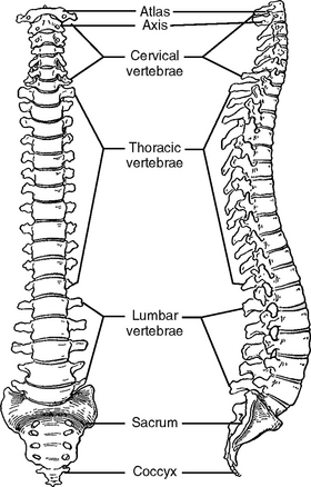


All of these parts contribute to the maintenance of a proper balance, which keeps the body erect, supports the head (which weighs 10 to 15 pounds), and holds the internal organs in an anatomically correct position. The muscular system of the trunk and its fascia ensure the stability of the erect spine and the range of motion of the spine during various physical activities. Co-activation of both flexor and extensor muscles along with a ligamentous structure is a required condition for securing the stable activity of the entire spinal area. Small muscles, tendons, and joint capsules serve as local segmental stabilizers between every two vertebrae.


All of the above mentioned muscles are vital for stabilizing the static and kinetic functions of the spine. A stabilized spine guarantees a smooth, accurate, and rhythmic functioning of the entire body during physical activities. For instance, the incredibly versatile design of the spine enables a gymnast to bend into almost unimaginable positions while a weight lifter can lift hundreds of pounds.


The spine is also in close interaction with the internal organs, and can be deformed by diseases of the internal organs, such as the lung, stomach, heart, or liver. These diseases disturb the balance of the mechanical support system and result in pain of the back. Likewise, an over-kyphotic spine will cause malfunction of the internal organs, especially the lungs. Thus, there is a close pathophysiologic relationship between the mechanical structure of the spine and the internal organs.


Numerous types of neck and lower back pain are triggered by mechanical abnormalities or injuries, inflamed soft tissues, or degenerative diseases, and of course, the pain can be idiopathic, or “origin unknown.” Please note that the INMAS protocol (see Chapter 5) can be used successfully for pain management regardless of which underlying causes produce the pain symptoms as long as the symptoms are physiologically recoverable.


The Foundation of the Spine: The Coccyx and the Sacrum


An analogy between the spine and a big tree will help to illustrate why many patients experience pain in this area. The trunk of a big tree (like the spine) has to support the canopy (the head) and numerous heavy branches (the upper limbs, ribs, and internal organs) that sprout from the trunk. This construction subjects the trunk to continuous mechanical stress and eventually the stress will affect the root system (the coccyx and sacrum bones).


The root system must be very strong and practically immovable to give adequate stability to the trunk and heavy branches of the tree. In calm weather, the tree ensures its symmetrical balance by having its branches spread in all directions and by evenly distributing gravitational stress among all the roots, which also spread in all directions. When a strong wind tilts the tree to one side, the symmetrical balance is lost and the roots from the opposite side of the tree sustain huge stress, which is many times higher than the weight of the tree (Figure 9-2). Using the physical principle of leverage, we can calculate the stress applied to the root system on the affected side of the tree. For example, if a tree weighs 500 pounds at its center of gravity, the affected part of the root system will sustain a stress of more than 5000 pounds.


The tree analogy explains why the foundation of the spine—the coccyx and sacrum—is so easily stressed and liable to be painful. Bending and twisting in an asymmetric manner are the predominant causes of lower back injury. The leverage described in the analogy with the tree is also applicable to our spine. For example, when a mother bends forward to pick up a 30-pound baby with both hands symmetrically, the stress on the spine and its foundation (sacrum and coccyx) could be 300 pounds. If the mother uses a correct bending posture, such as bending the knee first and staying close to the baby, the stress can be reduced to 100 pounds, which is distributed evenly between the muscles and joints on both sides of the body.


If our right hand picks up a 30-pound package (asymmetric action), the stress on the left sacroiliac joint and lumbar muscles on the left side can be more than 300 pounds, which will be distributed along the whole spine from the neck to the lower back. These are examples of the kind of potentially damaging stressful situations that our spine is exposed to daily, without our conscious awareness, in the workplace, at home, and during sports activities.


To correctly apply INMAS to the coccyx and the sacrum, an understanding of the anatomic structure of this area is important. The coccyx, which is also called the tailbone, is located at the base of the back. It consists of three to five fused bones. If we lose our balance and fall, the coccyx can break and a condition called coccygodynia may occur. We will discuss coccygodynia later in this chapter.


Above the coccyx are five more fused bones, the sacrum (S1-S5). The sacrum is a spade-shaped structure that is fixed between the two halves of the pelvis, which is the only structure that connects the spine to the lower limbs. Referring to the tree analogy, the two fused, relatively immovable bones—the coccyx and the sacrum—represent the roots of the tree. The two bones called ilium on either side of the pelvis make a joint on either side of the sacrum, and these are called the sacroiliac joints.


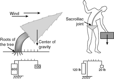
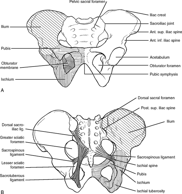
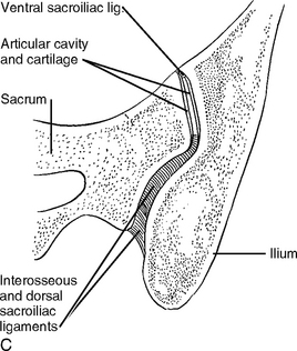
The sacroiliac (SI) joints differ from other joints. They are thicker and stronger and can move only a few millimeters (Figure 9-3). This relative immobility of SI joints stabilizes the spine against gravitational forces. The SI joints are also subject to the tremendous stresses and strains created by the forces of asymmetric imbalance as discussed in the above examples. The SI joints have to support the trunk, shoulders, arms, and head and to ensure a full range of motion.


During locomotive activity such as walking, running, carrying weights, raising the arms, or dancing, the joints transfer weight from the spine to the hip bones. The pathological effect of excessive or overextended stress will accumulate in the joints and result in soft tissue disease.


These joints are innervated by the lower lumbar and sacral nerves, as a result of which pathological conditions in the joints can produce lower back pain and sciatica. For example, when the SI joints are inflamed, movement of the spine in any direction causes pain in the lumbosacral part of the spine. Usually the pain is more pronounced at the limit of forward flexion, since the hamstring muscles hold the hip bones in a relatively fixed position while the sacrum is rotating forward as the spine bends down.


INMAS is effective in treating painful symptoms related to the SI joint region. Most SI joint pain is caused by inflammation of the soft tissues such as nerves and ligaments. When needles are inserted into inflamed tissues, the needle-induced lesions break the blood vessels, thus stimulating the local defense immune reaction. As a result of the immune reaction, inflammation of the soft tissues of the SI joint is reduced. The needle-induced secretion of endorphins from the spinal cord and the brain reduces the physiological stress caused by the joint pain, thereby accelerating tissue healing. Eventually, this process restores normal SI joint function. The degree of recovery of SI joint function depends on the degree of the injury and the self-healing capability of the body.


The INMAS protocol for treating SI joints is provided at the end of this chapter.


Lumbar Spine (L1-L5)


The lumbar part of the spine is situated above the sacrum. The lumbar spine has five large vertebrae, and each vertebra has two upper facets (superior articular facets) that emerge from the top and two lower facets (inferior articular facets) that descend from the bottom (Figure 9-4).
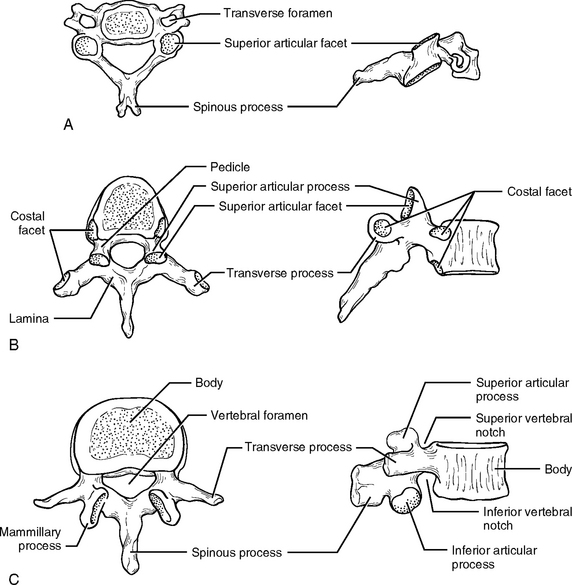


The functional unit of the lumbar spine consists of two adjacent vertebrae with an interposed intervertebral disk. The disk is a hydrodynamic structure that permits weight bearing and ensures the mobility of the unit itself and the mobility of the entire vertebral column. The two inferior facets of the vertebra above are loosely joined to the two superior facets of the vertebra below. During forward-and-back motion such as in fast dancing, the facets slide across each other. A side view shows that the lumbar spine is concave relative to the back of the body, and this is termed the lordotic curve.


The lumbar spine is located between two different and functionally conflicting parts of the spine and therefore is always subjected to substantial stress. Below the lumbar spine is the immobilized stability of the sacrum and above it are the heavy but flexible structures of the thoracic and cervical spine, which are required to be able to maintain a full range of spontaneous and rapid movement. This is why the lumbar spine is constantly engaged in trying to mediate an acceptable compromise between two conflicting functions, providing enough of both strength and flexibility at the same time.


Additional stress on the lumbar spine is created when a person holds weight in their hands with the arms extending away from the center of gravity of the body. In an upright posture the lumbar spine is supporting the axial compressive forces from the weight of the head, which is about 10 to 15 pounds, the weight of the upper limbs, and any additional loads that the upper limbs are carrying. When this compressive force is greater than half the body weight, particularly, the spine becomes unstable.


Because of these factors the lumbar spine is subject to a greater amount of constant stress than any other part of the spine, due to:


Cervical Spine


The cervical spine has seven vertebrae. These vertebrae get progressively smaller as they approach the bottom of the skull (see Figure 9-4). The cervical spine is the upper part of the spine and is capable of a great range of motion, in contrast to the foundation of the spine, the sacrum and coccyx, which has fused bones and almost immovable sacroiliac joints. For instance, the cervical spine allows the neck to turn 90 degrees in either direction, the ear to almost touch the shoulder, and the head to lean backwards more than 70 degrees.


The cervical spine differs from other parts of the spine in its agile motion and its ability to perform other important tasks; for instance, in addition to supporting the head, the neck provides a passage for air, food, nerves, and blood vessels.


The cervical spine is composed of two major complexes: the upper cervical segment (C1 and C2) and the lower cervical segment (C3 to C7). The occipital bone of the skull sits on the ring-shaped bone C1, which is called the atlas. The joint between them (0-C1), which is the first joint of the cervical spine, is called the atlantooccipital (AO) joint. This joint provides a very important function by anchoring the skull to the spine, and it is therefore necessarily the most immobile joint of the neck spine.


The second cervical vertebra, the axis, is the strongest of the cervical vertebrae. C2 has two large horizontal flat bearing surfaces called the superior articular facets. The atlas, C1, bearing the skull on top of it, rotates on these articular facets.


The distinctive feature of C2 is a blunt toothlike process called the odontoid process, or dens, projecting superiorly from the body (Figure 9-5). C1 uses the dens of C2 as its pivot for rotation. The front inner surface of the ring-shaped C1 is attached to the dens of C2, allowing C1 with the skull on top of it to rotate against the dens.
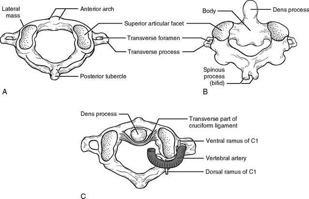


The joint between C1 and C2, called the atlantoaxial (AA) joint, is the most mobile part of the spine. Fifty percent of all cervical rotation occurs at the AA joint.


The first two vertebral joints differ from all other vertebral joints because their main function is to allow head rotation. C1 and C2 are jointed by ligamentous integrity, and not by bony facets as are all other typical vertebrae.


Among all vertebral joints the AO joint is the most immobile, whereas the AA joint is the most mobile. This functional characteristic makes the AA joint more vulnerable to wear and susceptible to pathological conditions like inflammation and arthritis.


The use of homeostatic acupoints (HAs) to implement functional restoration, as well as reducing or eliminating pain, makes it an effective modality in reducing inflammation, rebuilding the function of the joints, and relieving and controlling related pain syndromes.


The facet joint between C2 and C3 is a transitional structure located between the rotation joint above C3 and the flexion-extension joints below C3. The cervical spine from C3 to C7 is organized such that it can rotate and simultaneously flex forward or laterally, or extend backward. The cervical vertebrae have to maintain enough stability to support the head, which weighs 10 to 15 pounds, while providing enough flexibility to perform these motions.


The peripheral nerve network in the neck region is some of the most complicated nervous wiring in the body. Eight cervical nerves emerge from the intervertebral foramen to form the cervical plexus from C1 to C4, and brachial plexus from C5 to T1. The intervertebral foramen is the passage from which the spinal nerve emerges. The neck is also the passage for the autonomic nervous system, which balances the physiological activities of the majority of the internal organs. Thus, pathological neck problems will affect not only the head and the arms but many different organs ranging from the brain to the large intestine.


A functional unit of the spine consists of two neighboring vertebrae: one above and one below. Examination of what happens to a typical cervical functional unit during a car accident will help to explain how excessive motion of the neck or exposure to sudden external forces can cause neck problems and pain.


In a car accident a sudden external force causes the head to bend forward (forward flexion) and the upper vertebra slides forward about an axis of rotation. The anterior intervertebral disk space narrows, the posterior intervertebral disk space widens, and the intervertebral disk deforms. As a result of this excessive narrowing of the anterior intervertebral space, there may be damage to the cartilage of the anterior joint surface, and the intervertebral disk may be broken and pushed backward. The widened posterior joint may break the joint capsule or even cause tearing of the ligaments. The broken intervertebral disk becomes herniated and the damaged joint surface produces pain and can cause arthritis in the future.


Rotation of the cervical spine is a coupling motion that involves both rotation and lateral flexion. For example, when the head turns to the left, the upper vertebra rotates to the left while its left intervertebral disk space narrows and its right intervertebral disk space widens. When this rotation is caused by violent force, the sudden closure of the left intervertebral disk space will cause damage to the cartilage of the joints, break the intervertebral disk, and force it to the right. Rotation caused by violent force also may cause injury to the spinal nerves: the left intervertebral foramen becomes smaller and pinches the left spinal nerve, and the broken intervertebral disk bulges to the right and pushes the right spinal nerve.


During a whiplash accident, the horizontal force from the back causes the neck to overextend and overflex (Figure 9-6), which causes damage to both posterior and anterior joints. In addition, the translational back-and-forward movement of the upper vertebra closes the vertebral canal, which causes injury to the spinal cord. Understanding the basic mechanical structure of the spine and the nature of pathological damage sustained by the spine during an accident is the key to effective acupuncture treatment for back pain.
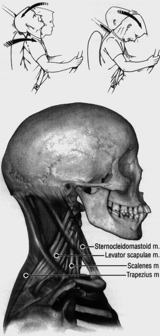


The INMAS protocol for treating whiplash will be provided in the end of this chapter. Please note that in the case of whiplash, pain symptoms such as neck pain, lower back pain, and upper back pain are all closely related because functionally and anatomically all three parts of the spine—the neck, the upper back, and the lower back—are interrelated structures that should be understood as one whole entity and treated as such.


All the components of the back are so dependent upon each other that a problem in one structure can damage all the others. For example, a mechanical neck injury causes inflammation in spinal joints, which will strain muscles, and chronic muscle strain can contribute to arthritis in the spinal joints. Neck pain and back pain are clinically coupled.


Muscle strain can lead to disk displacement, and displaced disks can further strain the muscles. Motions such as excessive forward flexion, excessive backward extension, or excessive lateral rotation cause injuries to bony joints that involve cartilages, disks, capsules, and ligaments. These injuries strain the muscles, and strained muscles irritate the spinal nerves. All these pathological changes to the basic mechanical structure of the spine produce pain, and eventually this pain can trigger chronic problems such as arthritis and degenerative diseases.


MUSCLE


Strained, overworked muscles are, in up to 80% of cases, the leading cause of all acute and chronic back pain, especially in the lower back.


Muscle is the most dynamic of all the mechanical components of the body. Muscles create energy and are the indispensable providers of the mechanical leverage that moves the bones. Thus, muscles are always in need of sufficient blood circulation to supply energy for movement and to eliminate toxic wastes from energy metabolism.


There is a widespread misconception that the spine is a very strong structure because, for example, a weight lifter can lift hundreds of pounds on his shoulders and in some cultures women habitually carry 40 to 50 pounds of cargo on their heads. In fact, the spine itself can carry only about 5 pounds and most of the work attributed to the spine is performed by the back muscles. The back muscles in turn are helped by the tendons and the ligaments to stabilize the spine and to allow the body to perform a variety of hard physical tasks.


Muscles perform at least three major mechanical functions:


Weak back muscles are not able to adequately perform the following functions:


Inadequate performance of these tasks leads to body weight imbalance or misplacement of the jointed bones, which will result in pain and the onset of tissue inflammation. Weak muscles are not able to support any sudden twist, or sudden change in posture, and will allow a disk to slip out of place, which in its turn can lead to disk rupture and neck and lower back pain syndromes.


Muscles are in a state of minimal contraction when the body posture is erect and static. The spine is maintained erect by muscles, fascia of the muscles, intervertebral disks, ligaments, and joint capsules. All the muscles are covered by a fascial sheath. The erect spine is primarily stabilized by the combined effort of these structures.


When the back muscles as well as the abdominal muscles have to be slightly contracted to maintain erect body posture, the fascial sheaths become taut. Neck and back problems happen when any one of the above mentioned structures experiences fatigue or injury and thus becomes a weak link in the chain.


Strain is a state of overstretching or overexertion of the musculature due to the application of excessive force. Strain disrupts the normal alignment of muscle fibers, ligaments, tendons, joint coverings, and the joints themselves. When strained, these tissues are subjected to microscopic tears that result in pain, bleeding, or inflammation and swelling.


Muscle strain may cause muscle spasms. A spasm is an uncontrollable intensive contraction of a muscle or group of muscles. A spasm increases tension with or without shortening of a muscle. A locally affected strained muscle can cause other muscles to become tight or to go into spasm in order to prevent an injury from spreading from the affected point to other structures.


Usually a spasm has no serious consequence and the pathological effect is not longlasting. However, when a tear caused by strain or spasm is severe or repeated, scar tissue is formed, which weakens the muscles and irritates the nerves, thus provoking pain syndromes. Acupuncture needling is very effective in the treatment of strained tissues and muscle spasms, especially at the acute phase.


A strained muscle or a muscle under spasm undergoes the vicious circle of energy crisis. Injured muscles become tightened. This tightness puts physical pressure on the blood vessels, which results in poor blood circulation. Insufficient circulation results in an insufficient supply of nutrients and oxygen being delivered to the injured muscles. Thus, the energy crisis begins.


As a result of energy deprivation, muscles become tighter and tighter. Then the nerves surrounded by the tightened muscles are subjected to hypoxia (low oxygen) and undernutrition and experience an energy crisis as well. During the energy crisis, metabolic toxins are accumulated in the injured muscles. This vicious circle of energy shortage creates muscle pain.


When needles are inserted into tight, strained, or spasmodic muscles, the needles push the tissues aside and create a microlesion. This process also stimulates a reflex reaction from the motor nerves, which relaxes the tight muscles and manifests as muscle twitching. Once the muscle starts to relax, adequate blood circulation to the injured muscle is restored, the vicious circle is broken, and the energy crisis is resolved. Improved blood circulation brings sufficient supplies of oxygen and nutrients and allows elimination of metabolic toxins. Thus, the muscles and nerves start the process of self-healing.


When the fibers of muscles, tendons, ligaments, or joint capsules are forcibly pulled and actually tear, then strain turns into sprain. A sprain can be a very serious and disabling physical condition. Sprains heal slowly and have a tendency to recur and become habitual. Acupuncture treatment will greatly speed up healing and reduce swelling at any stage, especially at the acute phase of the sprain. This is due to the way that needling and its lesions relax the injured muscles, which are usually tight, swollen, and deprived of nutrition and oxygen, with consequent vasoconstriction. Once the muscles are relaxed, normal blood circulation resumes, which brings the muscle physiology to normal condition. This normalization manifests itself by increased supply of nutrition and oxygen, reduced metabolic wastes, digestion of injured cells, and regeneration of new tissues. In addition, the needling stimulates the central nervous system to secrete neurochemicals such as endorphins, which improve cardiovascular function, and instigate immune reactions, which activate and accelerate the self-healing process.


The degree of muscle involvement may determine how an event is categorized, as a primary or a secondary cause of pain. “Primary muscle pain” means that the pathological condition of the muscles is the major cause of pain. Acupuncture is very effective for treating this type of pain. In cases of acute pain, relief can be achieved almost immediately after the beginning of treatment. In case of chronic pain, more treatments are needed for pain relief. “Secondary muscle pain” means that although the muscles are painful and in spasm, the condition is caused not directly by muscle injuries but by other injured structures such as nerves, joints, or disks, which contribute to the painful symptoms. For example, in the case of arthritis, muscle pain is caused by inflamed joints. More acupuncture treatments over a longer period of time are required to relieve secondary muscle pain because relief depends on healing the primary pain source, such as nerves, joints, disks, or other soft tissues.


Our basic protocol (INMAS) is used with individualized variations to treat all kinds of muscle pain and is provided at the end of this chapter.


As previously mentioned, the front muscles act as antagonists to the back muscles, which means that the front muscles flex the spine, while the back muscles extend it. These forces balance each other in order to maintain stability of the trunk and to secure various kinetic functions of the spine. When the front muscles are fatigued or injured, this balance is disturbed and the ability of the spine to maintain stability and proper posture is diminished. The tired front muscles became shortened and resistant to stretching, forming tender points within the muscle tissue, forcing the back muscles to stretch excessively and preventing relaxation of the back muscles.


Disturbance of this antagonistic balance causes neck, upper back, and lower back pain. This is why in the case of back pain symptoms it is important to carefully examine and to treat the front muscles without delay, namely the pectoralis major and pectoralis minor muscles on the chest as well as the external oblique, the anterior oblique abdominal, the transversus abdominis, and the rectus abdominis muscles.


Weak abdominal muscles often result in back pain. The abdominal muscles do much of the work when lifting or carrying loads, and so when weak abdominal muscles try to relieve the strain on back muscles during such activities, they will sustain further damage. This is why the abdominal muscles should be examined and needled in clinical acupuncture practice when a patient presents with back pain.


The physical condition of the limbs also influences the function of the spine. Thus it is especially important to maintain the flexibility of the powerful hamstring muscle in the back of the leg. When this strong muscle becomes tight, it limits the range of motion of the pelvis, and this can strain the lower back muscles and joints. In cases of back pain, the muscles on the limbs, especially the lower limb muscles of the thighs and legs, should be examined and their tightness relieved by acupuncture, which is a very effective treatment for it. Please note that a relaxed hamstring muscle permits a normal physiologic range of motion of the pelvis and thus allows coordinated movements between the pelvis and all other back muscles.


The Shortened Muscle Syndrome


Muscles become shortened for protective reasons, to prevent further damage to healthy muscle tissue when it is subjected to pathologic factors, such as cold, fatigue, repetitive overuse or overstretching, low oxygen, or deficient blood circulation. When a muscle becomes shortened a domino effect is initiated that can generate many painful conditions. Muscular shortening gives rise to tension in the tissues by pulling tendons and their attachments and by offering resistance to healthy stretching. Thus, the shortened muscles and the stressed tendons and attachments eventually cause such symptoms as neck and back pain, epicondylitis, tendinitis, and tenosynovitis. All of these symptoms share the same etiology, although they appear dissimilar and occur at different anatomic sites.


Shortened muscles limit the range of motion of joints. For instance, a pathological condition called “frozen shoulders” is a result of shortening of all the muscles around the shoulder joint in addition to a capsular problem like inflammation. The shortened muscles increase pressure on the articular surface of a joint, which results in joint pain (arthralgia). Muscle shortening is also often responsible for joint misalignment. For example, the shortened muscle (extensor hallucis longus) of the foot causes angulation of the great toe (hallux vulgus) and a painful bunion. Muscle shortening contributes to pathologic conditions of joints, such as restriction of the range of motion and misalignment, and eventually leads to degenerative arthritis and osteoarthritis. Shortened muscles also put pressure on the nerves and produce an entrapment syndrome. For example, carpal tunnel syndrome is a result of shortening in the pronator teres or pronator quadratus muscles.


The paraspinal muscles, when shortened, draw two adjacent vertebrae closer together, which narrows the intervertebral space and foramen (Figure 9-7). The results are a bulging disk and a compressed nerve root. A vicious circle is gradually built up: shortened muscles cause compressed nerve roots (radiculopathy), and compressed nerve roots lead to further muscle shortening.4
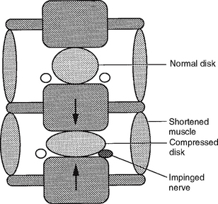


Acupuncture treatments can break this vicious circle by using needles to produce a minimal tissue injury, which stimulates the relaxation of muscle stress. The primary goal in the treatment of muscle shortening is relaxation of the affected muscle, and acupuncture needling achieves this goal more swiftly and precisely than any other medical modality. When a fine acupuncture needle pierces a muscle, it pushes aside tissue, disrupts the cell membrane, and inflicts a minute tissue injury, which mechanically creates a brief outburst of microcurrents (injury potentials). The microinjury that results from needling generates relatively longlasting currents that stimulate the mechanism of repair and regeneration of the affected tissue (see Chapter 3). Thus, acupuncture treatment eliminates the pathological condition responsible for muscle shortening. After treatment a patient feels either no pain or a significant alleviation of pain.


This mechanism explains the efficacy of acupuncture treatment in addressing pain syndromes resulting from radiculopathy or pathological conditions of the joints such as osteoarthritis.


NERVE TISSUE


The spinal cord is an extension of the brain and contains low-level nerve centers that process some pain sensation. The spinal cord sends out three kinds of nerves into the body: sensory, motor, and autonomic. Sensory nerves bring signals from the skin, muscles, tendons, joints, blood vessels, and organs to the spinal cord and then to the brain. These signals provide vital information about our activities and changes in the outside environment. Pain is the primary response to damage or irritation of nervous tissue.


Motor nerves transmit impulses originating in the brain and spinal cord down to the muscles of the skeleton and inner organs and to the glands in order to initiate and maintain appropriate movement. Autonomic nerves bring physiologic signals to the blood vessels and the internal organs to regulate their physiologic activities.


A nerve bundle may contain only one kind of nerve (sensory or motor nerves) or two or three kinds of nerves (mixed nerves). Normally sensory, motor, and autonomic nerves are interdependent because neither is able to function without the other. When nerves in the spinal cord have been disturbed by tissue damage—such as a pull, bruise, tear, swelling, or inflammation—they can trigger pain almost anywhere in the body.


Injury to nerves leads either to an involuntary, intense contraction (spasm) of the muscles in the body, the blood vessels, and organs, or to weakness or paralysis of the muscles as a result of insufficient contraction. A spasm causes overstimulation of sensory nerves and undernourishment of muscles because the tightness constricts blood vessels and interrupts the mechanism of nourishment. Weak muscles can easily be overstretched and thus be susceptible to painful tissue damage because this pathological overstretching releases chemicals that irritate the muscles (Chapter 3). Some of the pathophysiologic conditions of the spinal cord are discussed below.


Spinal Stenosis


Lumbar spinal stenosis is defined as a condition involving any type of narrowing of the spinal canal, nerve root canal, or tunnels of intervertebral foramina.5 Spinal stenosis can be either congenital or acquired. Acquired stenosis may be due to degenerative conditions (spondylolisthesis, see below), failed medical procedures (postlaminectomy, postfusion, postchemonucleolysis), or posttraumatic injuries, or it may be secondary to disk herniation (see below). Narrowing can occur in one or several locations of the same vertebral segment or it can affect several segments. The canal space can be narrowed by pathological changes in the soft tissues, scar tissue, or bony tissue impingement (bone spurs). Severe stenosis results in nerve compression. Soft tissue encroachment or abnormal bone growth such as osteophyte formation reduces the size of the intervertebral foramen and causes foraminal stenosis.

Only gold members can continue reading. Log In or Register to continue

Stay updated, free articles. Join our Telegram channel

Jun 11, 2016 | Posted by in BIOCHEMISTRY | Comments Off on 9: Back Pain: Neck, Upper Back, and Lower Back

Full access? Get Clinical Tree

Get Clinical Tree app for offline access