8

8 CASE 8


A 68-year-old man comes to his primary care physician complaining of tiredness and difficulty sleeping.


Six months earlier, the patient experienced myocardial infarction of the anterior wall of the left ventricle. A stent was placed in the occluded coronary artery, reestablishing perfusion. The patient now complains of difficulty sleeping at night, awakening with shortness of breath. He reports that he is able to sleep better using pillows to elevate his chest and head. The moderate exercise regimen that he followed after the myocardial infarction is now causing him to become progressively more short of breath.


PATHOPHYSIOLOGY OF KEY SYMPTOMS


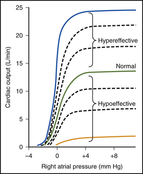
Blood entering the right atrium flows progressively through the right ventricle, pulmonary circulation, left atrium, and left ventricle before exiting the heart into the aorta. The ventricles are the power pumps in the heart. Ventricular output is determined by the preload on the ventricle and ventricular contractility.


The volume that flows through each of the cardiac chambers has to be equal or a fluid imbalance develops. This balance is achieved by the effect of preload on ventricular pumping. If the left ventricle pumps less blood than the right ventricle, the left ventricular end-diastolic volume will gradually increase. Left ventricular end-diastolic volume is “preload,” and an increase in preload will increase the pumping capability of the left ventricle. Left ventricular output increases, and the imbalance is corrected.


Echocardiography permits calculation of chamber size, chamber shape, wall thickness, and valvular function. This information can be used to calculate ejection fraction, which is the ratio of the stroke volume to the ventricular end-diastolic volume. The calculated left ventricular ejection fraction of 40% in this patient indicates that there is a systolic dysfunction of the left ventricle. Systolic dysfunction accounts for 60% to 70% of heart failure cases.


The presenting complaints of fatigue and nocturnal dyspnea are caused by impaired oxygen exchange at the alveolar capillaries. The impaired gas exchange is secondary to pulmonary edema, resulting from an impaired pumping ability of the left ventricle.


Myocardial infarction causes damage to the ventricular tissue and impairs the pumping ability. Because the infarct was in the left ventricle in this patient, left ventricular pumping is impaired. This results in a decreased arterial blood pressure and an elevated pressure in the left atrium and pulmonary circulation. The elevated volume in the left atria causes the appearance of a third heart sound owing to the turbulent flow of blood during atrial filling (Fig. 8-1).

< div class='tao-gold-member'>

Stay updated, free articles. Join our Telegram channel

Jul 4, 2016 | Posted by in PHYSIOLOGY | Comments Off on 8

Full access? Get Clinical Tree

Get Clinical Tree app for offline access