CASE 8
The mother of a 5-year-old boy, concerned about his inability to gain weight and his coughing and wheezing, appeared in the pediatrician’s office. The history revealed that the boy’s coughing and wheezing were worse at night and upon wakening and that his stools were foul smelling. The laboratory evaluation showed that sputum culture was positive for Pseudomonas aeruginosa and that the chloride sweat test was positive. Chest radiographs revealed hyperinflation, peribronchial cuffing, and mild bronchiectasis. A head x-ray showed opaque paranasal sinuses. The boy was diagnosed with cystic fibrosis. He was treated with gentamicin and piperacillin to eradicate the Pseudomonas aeruginosa, pancrelipase, and fat-soluble vitamins A, D, E, and K.
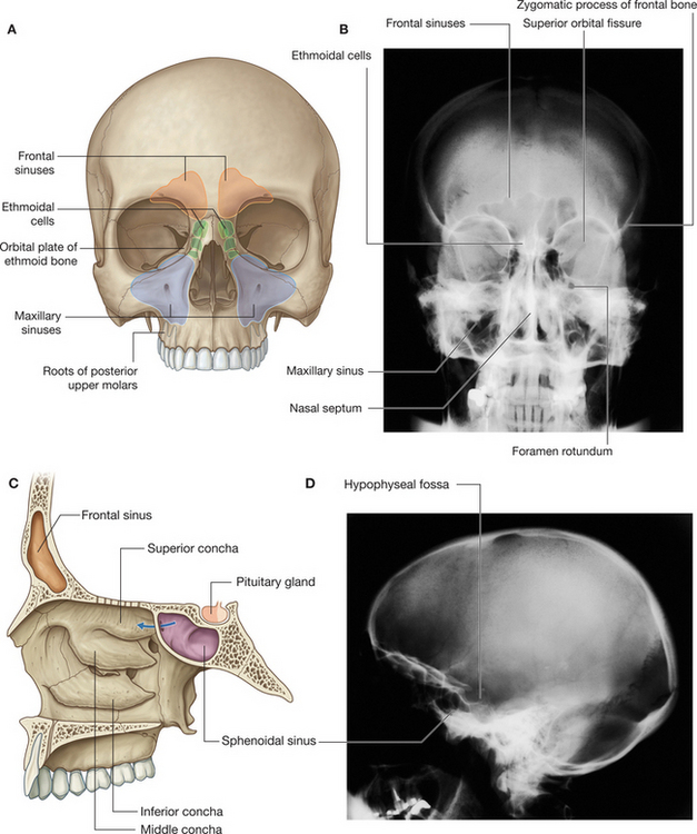
WHAT ARE THE PARANASAL SINUSES AND WHERE DO THEY DRAIN?
The paired, paranasal sinuses reside in the bones of the skull that surround the nasal cavity (Fig. 2-13). The sinuses are lined by ciliated, pseudostratified columnar epithelium containing goblet cells. The thickened mucus produced and secreted by the goblet cells contributes to their opaqueness in cystic fibrosis. The paranasal sinuses and their drainage are (Fig. 2-14):
Stay updated, free articles. Join our Telegram channel

Full access? Get Clinical Tree


