CASE 64
A 28-year-old Hispanic man was brought to the emergency department of a general hospital for severe headaches and two generalized seizures.
LABORATORY STUDIES
Imaging
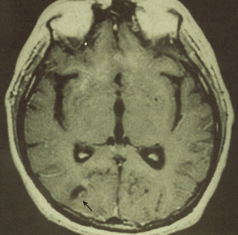
A CT scan of his brain revealed an intracranial calcified cyst, and further imaging with MRI confirmed the presence of similar lesions, some with a scolex visible (Fig. 64-1).
Diagnostic Work-Up
Table 64-1 lists the likely causes of illness (differential diagnosis). The presumptive diagnosis should be on the basis of clinical picture and epidemiologic information. The likelihood of exposure during foreign travel, and characteristic findings on CT or MRI scans, are adjuncts to clinical diagnosis. Lumbar puncture and peripheral blood collection are an essential beginning of investigation. Microbiologic investigation may include
TABLE 64-1 Differential Diagnosis and Rationale for Inclusion (consideration)
Stay updated, free articles. Join our Telegram channel
Full access? Get Clinical Tree
 Get Clinical Tree app for offline access

|