CASE 6
WHAT ARE THE NORMAL EVENTS IN THE SEPTATION OF THE PRIMITIVE ATRIUM?
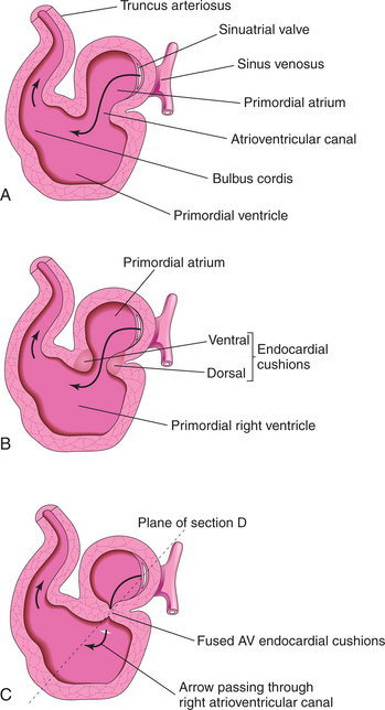
The dorsal and ventral endocardial cushions are masses of tissue that develop toward the end of the fourth week (Fig. 2-4). Growth of the cushions by the invasion of mesenchymal cells during the fifth week ultimately results in their fusion. This event divides the common atrioventricular canal into right and left atrioventricular canals. Moreover, fusion of the endocardial cushions is required for complete septation of the primitive atrium. The fused endocardial cushions ultimately become dense collagenous connective tissue of the cardiac skeleton.
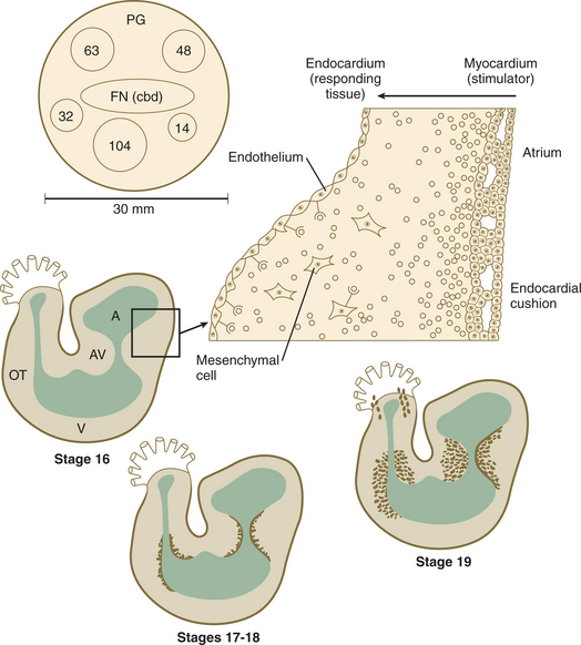
The formation of the endocardial cushions is orchestrated by an ensemble of molecules. The primitive myocardium induces the transformation of endothelial cells, which line the endocardium, into mesenchymal cells in the atrioventricular and outflow tract regions (Fig. 2-5). The inducible endothelial cells express the gene Msx-1. Endothelial cells that occupy other regions of the endocardium do not express Msx-1; therefore, they do not respond to the molecular inducer synthesized by the myocardium. The inducer, in part, is a molecular complex called adheron. Adherons, along with TGF-β1 and TGF-β3, are collectively responsible for the epithelial/mesenchymal transformation. In the absence of TGF-β1 and TGF-β3, the transformation of endothelial cells into mesenchymal cells fails to occur. The inducible endothelial cells also have to migrate from the endocardium into the cardiac jelly that occupies the space between the primitive myocardium and the endothelium. To migrate, the endothelial cells must decrease their synthesis of N-CAM, a protein that anchors the cells in place. The down regulation of N-CAM ultimately allows these cells to become motile. Once these motile cells have morphed into mesenchymal cells, they inactivate adherons through the secretion of proteases. These molecular events are also required for normal development of the heart valves.