CASE 55
WHAT STRUCTURE PRODUCES AQUEOUS HUMOR AND HOW IS THIS HUMOR DRAINED?
Located between the ora serrata and the margin of the lens is a wedge-shaped extension of choroid called the ciliary body (Fig. 7-20, upper left). Extending from the anterior part of the ciliary body toward the lens are numerous (approximately 70) ciliary processes. Attached to the ends of these processes and to the lens are the suspensory ligaments of the lens. Most of the ciliary body, except for the ciliary processes, is composed of smooth muscle forming the ciliary muscle. The ciliary muscle has three bundles of smooth muscle. The lateral-most muscle bundle, which abuts the sclera, stretches the choroid. This muscle, called the tensor muscle of the choroid and also known as the muscle of Brücke, facilitates drainage of aqueous humor. The other two muscle bundles are involved in accommodation for near vision. Contraction of these smooth muscle bundles paradoxically reduces the tension on the suspensory ligaments of the lens. The reduced tension increases the refractive power of the lens by increasing its thickness, thus enabling the person to view near objects. The core of the ciliary body is the vascular layer, which is composed of loose connective tissue containing elastic fibers and numerous blood vessels. The cores of the ciliary processes contain loose connective tissue supporting many fenestrated capillaries.
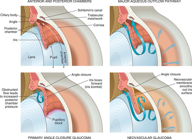
Aqueous humor is a relatively protein-free filtrate of plasma. The plasma is filtered by the fenestrated capillaries present in the ciliary processes. The filtrate is then transported into the pigmented columnar cells of the basal layer of ciliary epithelium and relayed to the nonpigmented cells of the superficial layer. From this layer, aqueous humor is released in the posterior chamber of the eye, which is located between the vitreous body and the iris. Aqueous humor then flows through the pupil of the iris to enter the anterior cavity located between the iris and the cornea. To maintain normal intraocular pressure, aqueous humor, which is being constantly produced, must be drained from the anterior chamber. The biological drainage apparatus is located near the limbus. The limbus is at the junction of the cornea and sclera. The apparatus is composed of a trabecular meshwork of endothelium-lined spaces that leads to the canal of Schlemm (sinus venosus sclerae). Aqueous humor filters through this meshwork before being collected by the canal of Schlemm (Fig. 7-20, upper right).


