CASE 52
WHAT CRANIAL NERVE IS INVOLVED AND WHAT IS ITS COURSE?
Bell’s palsy involves the facial nerve. The two divisions of the facial nerve (large motor root and a small intermediate nerve) exit the brainstem at the pontomedullary junction. The motor root is responsible for the innervation of the muscles of facial expression, and the intermediate nerve conveys gustatory, parasympathetic, and somatosensory fibers. The facial nerve leaves the cranial cavity by entering the internal acoustic meatus, traversing the facial canal in the petrous portion of the temporal bone, and exiting at the stylomastoid foramen (Fig. 7-9). Because of its long intraosseous journey, the facial nerve is vulnerable to compression within the facial canal.
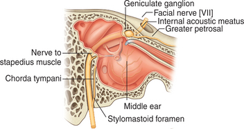
FIGURE 7-9 Facial nerve traversing facial canal of temporal bone.
(Drake R, Vogl W and Mitchell A: Gray’s Anatomy for Students. Churchill Livingstone, 2004. Fig. 8-124)
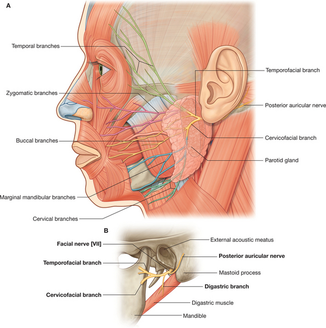
Once it exits the stylomastoid foramen, the facial nerve branches into the posterior auricular nerve and a main trunk (Fig. 7-10). The main trunk and the parotid plexus that it forms are embedded in the parotid gland. The plexus gives rise to the five terminal branches of the facial nerve: temporal, zygomatic, buccal, marginal mandibular, and cervical (Fig. 7-10). The distribution of the five terminal branches and the posterior auricular nerve are purely motor. Table 7-1 describes the muscular distribution of the facial nerve. In addition to the many muscles listed in the table, the facial nerve also innervates the stapedius, a small muscle in the middle ear.