CASE 51
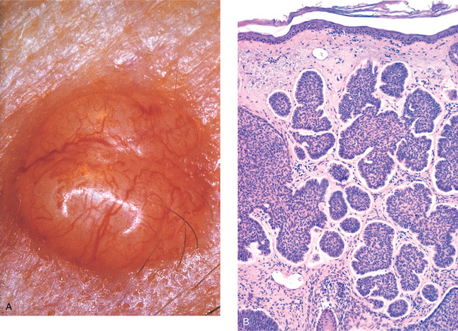
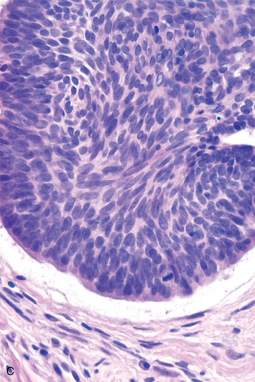
A 52-year-old white man presented to his family physician complaining of a bleeding growth on his nose. The patient states that his job requires him to frequently work outside and that he seldom uses protection against the sun even though he is of fair complexion. Visual examination of the growth showed that it was shiny and pink-colored with dilated blood vessels and a white rolled margin (Fig. 7-4A). The center of the papule was ulcerated and bleeding and measured 2 cm in diameter. Microscopic examination of a biopsy of the papule confirmed a diagnosis of basal cell carcinoma. The lesion was surgically excised.
HOW DOES SKIN FORM EMBRYOLOGICALLY?
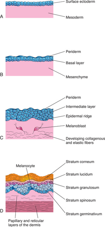
The skin is formed from two germ layers. The epidermis is derived from ectoderm, whereas the underlying dermis develops from mesoderm-derived mesenchyme. The basic events of development of the epidermis are (Fig. 7-5):

FIGURE 7-5 Stages of skin development. A, 4 weeks. B, 7 weeks. C, 11 weeks. D, Newborn.
(Moore K and Persaud TVN: The Developing Human, 7e. WB Saunders, 2003. Fig. 20-1.)
Most of the dermis differentiates from the lateral somatic mesoderm.
HOW IS SKIN ON THE NOSE DESCRIBED HISTOLOGICALLY?
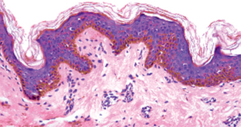
Based on the thickness of the epidermis, skin can be classified as thin or thick. In this case, the skin on the nose is categorized as thin skin. Microscopically, four layers can be observed in the epidermis of thin skin. From deep to superficial, these layers are (Fig. 7-6):

FIGURE 7-6 Histologic features of thin skin. Note the presence of melanin in the basal layer of the epidermis.
(Kerr J: Atlas of Functional Histology. Mosby, 1999. Fig. 8-3b.)
Stay updated, free articles. Join our Telegram channel

Full access? Get Clinical Tree


