CASE 50
ALZHEIMER’S DISEASE IS ASSOCIATED WITH A DECREASE IN CHOLINERGIC INPUT TO THE CEREBRAL CORTEX. WHAT NUCLEUS OF THE BRAIN CONTAINS THESE CEREBRAL CORTEX-PROTECTING, CHOLINERGIC NEURONS THAT DEGENERATE IN ALZHEIMER’S?
ALZHEIMER’S DISEASE IS ALSO ASSOCIATED WITH A LOSS OF NEURONS IN TWO REGIONS OF THE BRAIN INVOLVED IN THE FORMATION OF NEW MEMORIES AND LEARNING. WHAT ARE THESE REGIONS AND WHAT ARE THE CLASSIC MICROSCOPIC ABNORMALITIES THAT ARE OBSERVED IN THESE AREAS?
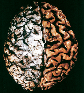
This global decrease in neurons results in gross pathologic changes, such as cerebral atrophy, widening of the sulci, and dilatation of the ventricles (Fig. 7-1).
Several derangements in microscopic structure are observed in Alzheimer brains:
The two pathologically classic abnormalities are neuritic plaques and neurofibrillary tangles. Neuritic plaques are focal aggregations of argyrophilic (silver-loving) dystrophic neurites (dystrophic neurites are thickened or irregular processes of neurons) surrounding a central core of amyloid (Fig. 7-2A, B
Stay updated, free articles. Join our Telegram channel

Full access? Get Clinical Tree


