CASE 5
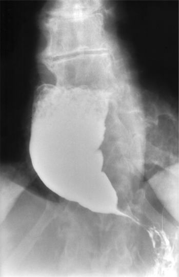
A 46-year-old woman presented to her physician complaining of dysphagia, regurgitation of undigested food and liquid and modest weight loss. The history revealed that the symptoms have lasted for 3 years and that she has a feeling of retrosternal fullness after meals. This feeling of fullness can be relieved at times by drinking a large glass of water and by standing with arms over her head. The radiographic imaging study (barium swallow and fluoroscopy) showed a dilated esophagus with a distinct air-fluid interface and a “bird’s beak” silhouette (Fig. 2-1) in the region of the lower esophageal sphincter (LES) and no significant peristalsis in the esophagus. Save for an inflamed esophageal mucosa, the endoscopic examination was unremarkable. Esophageal manometry showed incomplete relaxation of the LES and aperistalsis of the esophagus. The patient was diagnosed with primary achalasia (unknown etiology), which was successfully treated with pneumatic dilation of the LES.

FIGURE 2-1 Esophagogram showing the characteristic “bird’s beak” appearance of achalasia.
(Towsend C et al.: Sabiston Textbook of Surgery, 17e. WB Saunders, 2004. Fig. 39-11. Courtesy of Ronella A. Dubrow, M.D., MD Anderson Cancer Center, Houston, TX.)
WHAT IS THE BLOOD SUPPLY TO THE ESOPHAGUS?
The esophagus is richly supplied by arteries. These arteries are:
WHAT IS THE INNERVATION OF THE ESOPHAGUS?
The nerve supply to the esophagus is from the:
Stay updated, free articles. Join our Telegram channel

Full access? Get Clinical Tree


