CASE 38
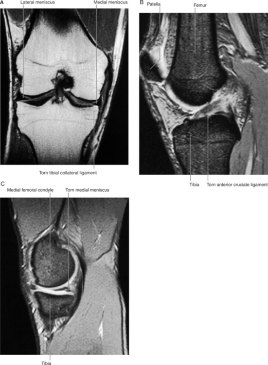
A 27-year-old linebacker was clipped on the posterolateral aspect of his knee during a professional football game. The player complained of severe pain in his right knee and stated that he heard his knee pop right after he was hit. The team physician immediately conducted a physical examination of the injured player’s knee and observed the following: (1) the player’s pain was exacerbated with medial rotation of the leg; (2) a positive anterior drawer sign was observed; and (3) abduction of the slightly flexed knee caused the medial side of the joint to separate more than normal. Several minutes later, the joint cavity of the knee swelled with blood (hemarthrosis). Imaging studies confirmed the diagnosis of the “unhappy triad” of the knee (Fig. 5-7). The player was then referred to an orthopedic surgeon for reconstructive knee surgery.
WHAT ARE THE THREE DAMAGED STRUCTURES THAT CONSTITUTE THE “UNHAPPY TRIAD”?
The “unhappy triad” is characterized by injury to the medial (tibial) collateral ligament, the anterior cruciate ligament, and the medial meniscus (Fig. 5-7).
WHAT ARE THE FUNCTIONS OF THE COLLATERAL LIGAMENTS, THE CRUCIATE LIGAMENTS, AND THE MENISCI?
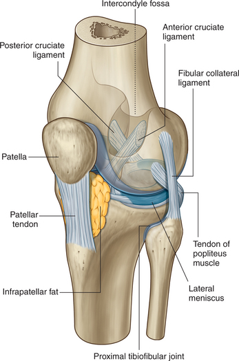
The lateral and medial collateral ligaments (Fig. 5-8) function to check extension and lateral rotation of the tibia, as well as limit side-to-side movements. Individually, the medial collateral ligament would limit abduction of the tibia, whereas its fellow would check adduction of the tibia. In the clinical scenario above, it was excessive abduction that caused the medial collateral ligament to rupture.

FIGURE 5-8 Knee joint with joint capsule removed.
(Drake R, Vogl W and Mitchell A: Gray’s Anatomy for Students. Churchill Livingstone, 2004. Fig. 6-68.)
The anterior and posterior cruciate ligaments (Figs. 5-8 and 5-9
Stay updated, free articles. Join our Telegram channel

Full access? Get Clinical Tree


