CASE 37
Concerned about the developmental delays in motor skills of their 5-year-old boy, the parents scheduled an appointment with a pediatrician. The parents informed the physician that their son seemed uncoordinated as he had difficulty running. The results of the physical examination detected hypertrophy of the calf muscles and diminished proximal tendon reflexes. Laboratory tests revealed a deletion of the dystrophin gene and elevated serum creatine kinase levels (40 times normal). Based on this information, the boy was diagnosed with Duchenne’s muscular dystrophy.
WHAT IS DYSTROPHIN AND WHAT IS ITS FUNCTION?
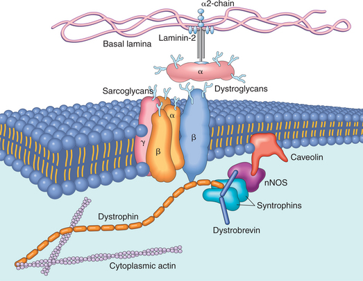
Dystrophin is an intracellular glycoprotein found in skeletal and cardiac muscle (Fig. 5-5) that links the microfilament actin to a group of transmembrane proteins (i.e., dystroglycans and sarcoglycans). The transmembrane proteins anchor the sarcolemma to the extracellular matrix. Functionally, dystrophin imparts structural integrity to the sarcolemma by forming a molecular interface that transmits forces from the contractile elements of the muscle cell to the extracellular matrix.