CASE 33
A 54-year-old African-American man presented to his physician with complaints of frequent urination and a decreased force of urination. The physician detected a dense nodular region of the prostate gland during a digital rectal examination. Furthermore, a blood test indicated that the patient’s prostate-specific antigen (PSA) was high (8 ng/mL). Based on these observations, a biopsy was obtained from the prostate gland and submitted to pathology for analysis. The pathology report confirmed the physician’s suspicion that his patient had prostate adenocarcinoma, a common form of prostate cancer.
WHAT STRUCTURES CAN BE PALPATED DURING A DIGITAL RECTAL EXAMINATION?
The structures that can be palpated during a digital rectal examination in males and females are identified in Table 4-2.
TABLE 4-2 Palpable Structures During A Digital Rectal Examination
| Male | Female |
|---|---|
| Penile bulb | Uterine cervix |
| Prostate gland | Pathological changes in the: |
| Seminal vesicles, if enlarged | Ovaries |
| Base of bladder, if distended | Uterine tubes |
| Inferior sacrum and coccyx | Broad ligaments |
| Ischial spines and tuberosities | Recto-uterine pouch |
| Internal iliac lymph nodes, if enlarged |
WHAT IS THE LYMPHATIC DRAINAGE OF THE PROSTATE GLAND?
Principal lymphatic drainage of the prostate is to the internal iliac, obturator, and sacral nodes.
HOW IS THE PROSTATE GLAND DESCRIBED HISTOLOGICALLY?
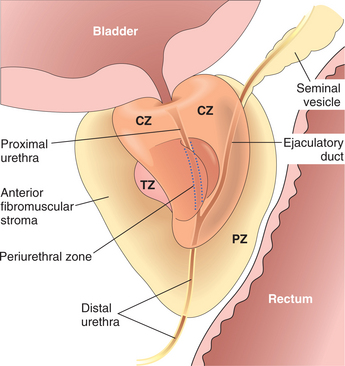
The prostate gland has an average mass of 20 g and is divided into fibromuscular and glandular regions. The fibromuscular region is located along the anterior border of the gland. The glandular component, which comprises two-thirds of the prostate, contains 30 to 50 compound tubuloalveolar glands that reside in three distinct histologic zones (Fig. 4-17):
Stay updated, free articles. Join our Telegram channel

Full access? Get Clinical Tree


