27

27 CASE 27


A 25-year-old woman comes to her primary care physician complaining of oral bleeding.


The patient has been on low molecular weight heparin for the past 2 years after the diagnosis of a pulmonary embolism.


PATHOPHYSIOLOGY OF KEY SYMPTOMS


The primary symptoms of this patient are all related to impaired platelet function. Platelets are usually capable of sealing small breaks in blood vessels without causing activation of the clotting cascade. When platelets encounter a damaged vascular endothelial cell, the platelets aggregate and form a platelet plug, sealing the damaged vessel. Impaired platelet function can cause prolonged bleeding, and the bleeding results in the appearance of small red dots on the skin and in the mouth (petechiae). It can also cause the appearance of somewhat larger or red purple dots (purpura) or even larger hemorrhagic bullae.


Platelets are fragments of megakaryocytes that play two roles in hemostasis. They form a platelet plug to seal small openings in blood vessels (temporary hemostasis) and contribute platelet factor III to the coagulation cascade. The formation of the fibrin clot helps to reinforce the platelet plug and to occlude larger areas of damage.


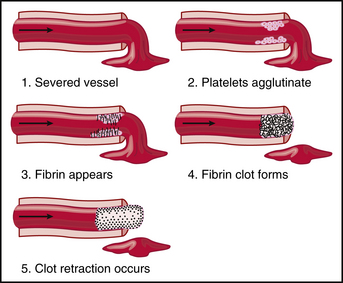
Damage to the endothelium of the blood vessel allows blood to come in contact with collagen. When a platelet contacts collagen, the platelet becomes activated and adheres to the damaged area. The platelets undergo a release reaction, secreting adenosine diphosphate (ADP), serotonin, and thromboxane. These substances cause the activation of adjacent platelets and allow the platelets to accumulate into a “platelet plug” (Fig. 27-1).

< div class='tao-gold-member'>

Stay updated, free articles. Join our Telegram channel

Jul 4, 2016 | Posted by in PHYSIOLOGY | Comments Off on 27

Full access? Get Clinical Tree

Get Clinical Tree app for offline access