CASE 22
A 23-year-old man was admitted to the hospital for fever, nonproductive cough, progressive shortness of breath, and fatigue for 2 weeks.
LABORATORY STUDIES
Imaging
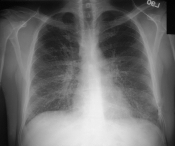
A chest radiograph revealed bilateral air-space consolidation with interstitial and alveolar markings (Fig. 22-1).
Diagnostic Work-Up
Table 22-1 lists the likely causes of illness (differential diagnosis). Sputum samples should be obtained for Gram and acid-fast stains and routine cultures to rule out the likely fungi (including P. jiroveci), mycobacteria, and other bacterial causes. Additional tests for specific microbiologic diagnosis may include methenamine silver stain or direct fluorescence antibody (DFA) stain of bronchoalveolar lavage (BAL) specimens.
TABLE 22-1 Differential Diagnosis and Rationale for Inclusion (consideration)
Rationale: A diagnosis of pneumonia should be considered. In an AIDS patient (CD4+ cell count <200/μL) who is not taking prophylaxis for opportunistic infections, the clinical presentation above is highly suggestive of P. jiroveci pneumonia. TB should always be considered in any HIV-positive patient with a respiratory syndrome, due to the greatly increased risk of TB in these patients. Other fungi (including Histoplasma capsulatum) and typical lower respiratory bacterial pathogens (including Streptococcus pneumoniae) are commonly seen. CMV and Nocardia are also likely to be considered, although CMV is usually seen with much lower CD counts (<50). The atypical respiratory bacterial pathogens (e.g., M. pneumoniae) and respiratory viruses (e.g., adenovirus) are less likely.
Stay updated, free articles. Join our Telegram channel

Full access? Get Clinical Tree


