CASE 22
A 54-year-old man presented to his physician with complaints of decreased urine output and dark-colored urine. His arterial pressure was 150/90. The results of urinalysis were positive for the presence of erythrocytes, leukocytes, and casts. It also showed that specific gravity was above normal and that proteins were elevated. The laboratory result for renin showed that its secretion was suppressed. The patient’s glomerular filtration rate, as measured by radionuclide clearance, was decreased. The patient was diagnosed with glomerulonephritis.
HOW IS THE RENAL CORPUSCLE DESCRIBED HISTOLOGICALLY?
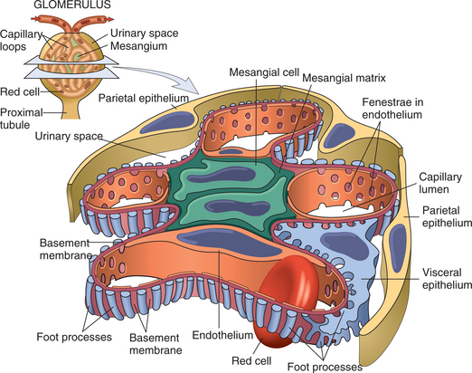
Each kidney contains 1 million to 4 million renal corpuscles. Renal corpuscles are composed of an outer epithelial capsule, which is called the parietal layer of the glomerular (Bowman’s) capsule (Fig. 3-28). Inside the capsule, a tuft of capillaries, the glomerulus, is covered by an inner epithelial layer, called the visceral layer of the glomerular capsule. The visceral layer is composed of cells called podocytes. The podocytes and glomerular capillaries constitute the filtration barrier (see below). The space between the parietal and visceral layers is the capsular (Bowman’s) space. This space receives the ultrafiltrate from the blood flowing through the glomerulus. The renal corpuscle possesses two poles: vascular and urinary. The vascular pole receives the afferent arteriole, which delivers arterial blood to the glomerulus, and an efferent arteriole, which conveys blood away from the glomerulus. The urinary pole is the end of the renal corpuscle that is continuous with the proximal convoluted tubule.

FIGURE 3-28 Illustration of a renal corpuscle.
(Kumar V, Abbas A and Fausto N: Robbins & Cotran Pathologic Basis of Disease, 7e. WB Saunders, 2004. Fig. 20-1.)


