CASE 20
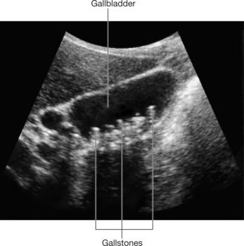
A 52-year-old woman presents in the emergency room with complaints of sudden right upper quadrant pain, nausea, and vomiting. The patient weighed 170 lb, height was 60 inches, and she had a mild fever. Tenderness and guarding inferior to the right costal margin and a positive Murphy’s sign were noted during the physical examination. Laboratory results showed slightly elevated levels of serum bilirubin, alkaline phosphatase, and leukocytes. Ultrasound imaging detected gallstones (Fig. 3-22) and a thickened wall of the gallbladder. The patient was diagnosed with acute calculous cholecystitis. She was placed on intravenous antibiotics and nonsteroidal analgesics to alleviate the pain. Laparoscopic cholecystectomy was performed 2 days later.

FIGURE 3-22 Ultrasound demonstrating stones in the gallbladder.
(Drake R, Vogl W and Mitchell A: Gray’s Anatomy for Students. Churchill Livingstone, 2004. Fig. 4-94.)
WHAT ARE THE BORDERS OF THE HEPATOCYSTIC TRIANGLE (OF CALOT)?
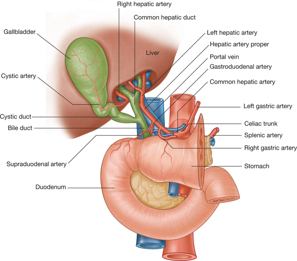
The hepatocystic triangle has three borders: superior, medial, and lateral. The borders are formed as follows (Fig. 3-23):

FIGURE 3-23 Triangle of Calot is bordered by the cystic artery, common hepatic duct, and cystic duct.
(Drake R, Vogl W and Mitchell A: Gray’s Anatomy for Students. Churchill Livingstone, 2004. Fig. 4-99.)


