CASE 16
A 52-year-old man presented to the physician with complaints of loss of appetite, weight loss, and weakness. The patient commented during the history that he drinks six to ten beers a day and more on weekends. The physical examination showed the following: jaundice, peripheral edema, distended abdomen, ascites, caput medusae, and wasted extremities. Laboratory tests revealed hypoproteinemia, hyperbilirubinemia, anemia, increased prothrombin time and elevated levels of serum aminotransferase and serum alkaline phosphatase. A liver biopsy confirmed the diagnosis of alcoholic cirrhosis.
WHERE ARE THE PORTOSYSTEMIC ANASTOMOSES AND WHAT RESULTS IF THEY BECOME DISTENDED?
When the pressure is normal in the hepatic portal system, all the blood following to the liver is delivered to the hepatic veins that empty into the inferior vena cava. However, when portal venous blood flow is obstructed, as in the later stage of alcoholic cirrhosis, the ensuing elevated portal pressure shunts blood into collateral veins that empty into the systemic venous circulation. The largest of these portosystemic anastomoses occur at the following locations (Fig. 3-9):
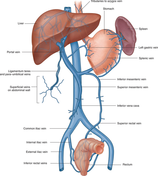
FIGURE 3-9 Portosystemic anastomoses.
(Drake R, Vogl W and Mitchell A: Gray’s Anatomy for Students. Churchill Livingstone, 2004. Fig. 4-106.)
These portosystemic anastomoses are further described in Table 3-2.
TABLE 3-2 Portosystemic Anastomoses
| Location | Description | Clinical Significance |
|---|---|---|
| Gastroesophageal | Esophageal veins that empty into the left gastric vein of the portal system anastomose with esophageal veins that drain into the azygos vein of the systemic venous system. | Esophageal varices form at the gastroesophageal junction. |
| Anorectal | Superior rectal vein, which empties into the portal system, anastomoses with the middle and inferior rectal veins. The middle and inferior rectal veins drain into the internal iliac and internal veins of the systemic venous system, respectively. | Hemorrhoids occur at the anorectal junction. |
| Paraumbilical | Paraumbilical veins, which accompany the ligamentum teres and median umbilical ligament, anastomose with subcutaneous veins of the anterior abdominal wall (e.g., superficial epigastric vein, which empties into the great saphenous vein). | Caput medusae forms around the umbilicus. |
| Retroperitoneal | Anastomoses occur between the veins of the bare area of the liver and veins of the diaphragm and right internal thoracic vein. |


