CASE 15
A 20-year-old woman presents to the emergency room with complaints of right lower abdominal pain, nausea, and loss of appetite. When questioned about the pain, she stated that it was general in nature. But since then it moved to just under the sternum (epigastrium) then to the belly button, and now the patient felt it in her right lower abdominal area. Examination of her abdomen revealed diminished bowel sounds and rebound tenderness with muscle spasms in the right lower abdominal quadrant. Rovsing’s, psoas, and obturator signs were also observed. Laboratory studies were unremarkable save for mild leukocytosis with increased neutrophils. The patient was diagnosed with appendicitis and a laparoscopic appendectomy was performed.
WHAT ARE ROVSING’S, PSOAS, AND OBTURATOR SIGNS?
The Rovsing’s, psoas, and obturator signs are described in Table 3-1.
TABLE 3-1 Description of Rovsing’s, Psoas, and Obturator Signs
| Sign | Description |
|---|---|
| Rovsing’s | Pressure on the left lower abdominal quadrant produces pain in the right lower quadrant. |
| Psoas | Extension of the right thigh stretches the psoas major muscle. The presence of pain during this procedure suggests an inflamed appendix contacting the stretched muscle. |
| Obturator | Passive medial rotation of the flexed thigh elicits pain when the inflamed appendix contacts the obturator internus muscle. |
WHAT IS MCBURNEY’S POINT AND WHERE IS IT LOCATED?
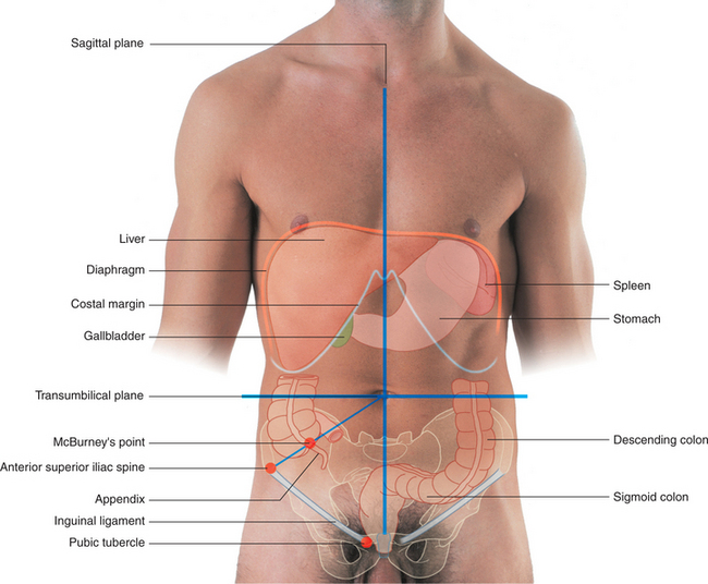
McBurney’s point is the surface landmark that indicates the approximate location of the appendix (Fig. 3-5). To locate McBurney’s point, a line can be drawn from the right anterior superior iliac spine to the umbilicus. McBurney’s point is one-third of the way along this line from the anterior superior iliac spine.
WHAT ARE THE LAYERS OF THE ABDOMINAL WALL THAT ARE INCISED OVER MCBURNEY’S POINT TO GAIN ACCESS TO THE ABDOMINAL CAVITY?
An incision of the abdominal wall over McBurney’s point would cut through the following layers (Fig. 3-6):
Stay updated, free articles. Join our Telegram channel

Full access? Get Clinical Tree


