Thoracic Surgery
LEARNING OBJECTIVES
After studying this chapter the reader will be able to:
• Correlate surgical anatomy and physiology of the thoracic cavity
• Discuss the purposes and potential complications and risks associated with thoracic surgery
• Discuss diagnostic methods for determination of surgical interventions and approaches
• Identify tissue layers involved in opening and closing thoracic incisions
• List the pharmacologic and hemostatic agents used during thoracic surgery
Overview
Thoracic surgery, like other specialties, has evolved with the development of surgical techniques and treatments, such as blood transfusion, anesthetic delivery, and screening procedures. During the past 50 years the understanding of pathophysiology and improved techniques have expanded the field of thoracic surgery. The thoracic specialty extends beyond the surgical arena into infectious disease, trauma, and oncology. Improved technology and the determination to treat diseases previously considered untreatable with operative and other invasive procedures continue to improve the recovery rate for patients experiencing thoracic diseases. As the ability to treat thoracic disease has improved, the responsibilities of the surgical team have expanded, resulting in accomplishments throughout the years that have provided an extensive knowledge base and specialized perioperative practitioners.
Surgical Anatomy
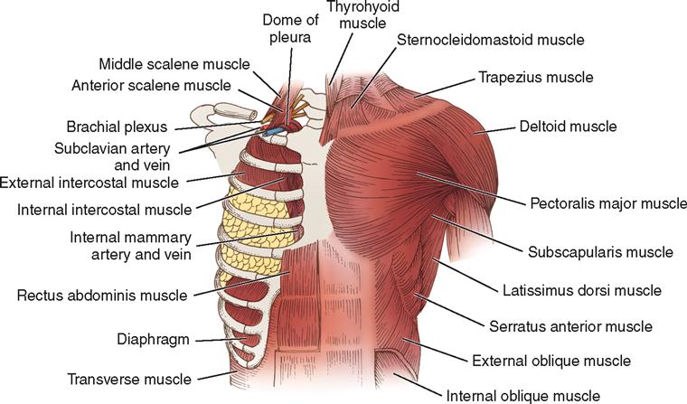
The skeletal framework of the thorax is formed anteriorly by the sternum and costal cartilages, laterally by the 12 pairs of ribs, and posteriorly by the 12 thoracic vertebrae (Figure 14-1). This airtight compartment is enclosed in the root of the neck by Sibson’s fascia and is separated from the abdomen by the diaphragm.
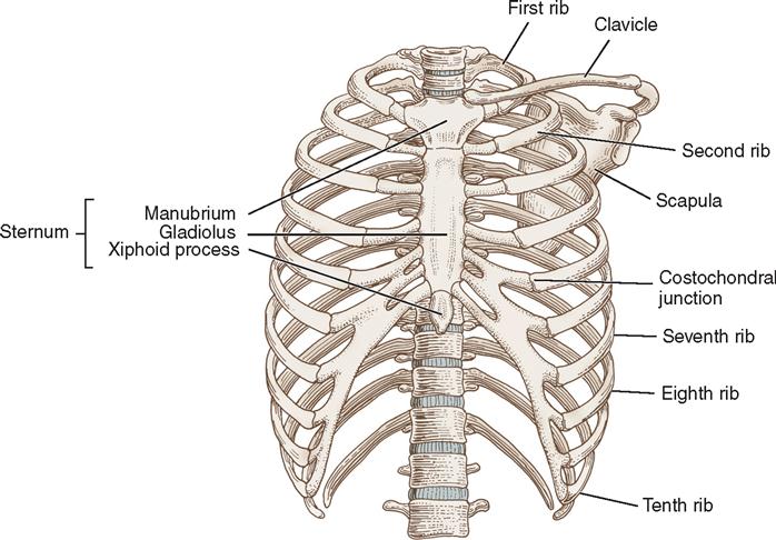
The sternum forms the anterior thoracic wall in the midline. It consists of three parts: (1) the upper part, or manubrium; (2) the body, or gladiolus; and (3) the lower cartilage, or xiphoid process. The manubrium articulates with the clavicles and the first two ribs on each side; the gladiolus articulates with the remaining true ribs by separate costal cartilages; and the xiphoid fuses with the gladiolus in early development and is attached to the diaphragm by the substernal ligament.
Normally the lateral walls of the thorax are formed by the 12 pairs of ribs. Posteriorly, each pair of ribs articulates with its corresponding thoracic vertebrae. Anteriorly, the first seven ribs articulate with the sternum. The eighth, ninth, and tenth ribs articulate with the costal cartilages of the rib above; however, the eleventh and twelfth ribs are not fixed to the costal arch (see Figure 14-1).
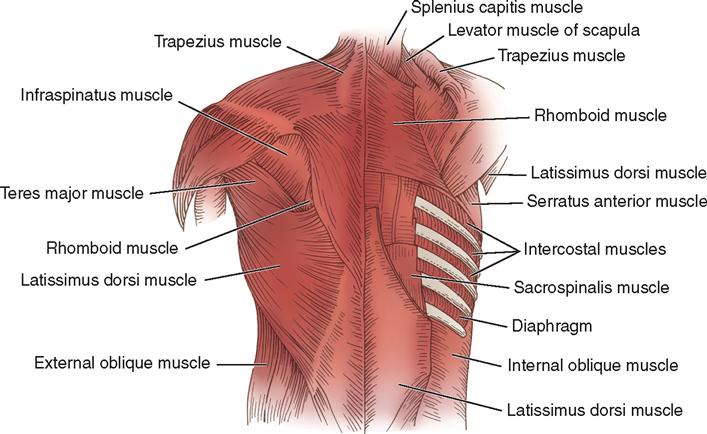
The muscles of each hemithorax (Figures 14-2 and 14-3) include the 11 external and 11 internal intercostal muscles, which fill the spaces between the ribs. An intercostal artery, vein, and nerve accompany each intercostal muscle. The arteries communicate with the internal thoracic artery anteriorly and arise from the aorta posteriorly. The intercostal veins follow the course of the arteries and communicate with the mammary veins anteriorly and with the azygos and hemiazygos veins posteriorly.
During surgery, great care is taken to prevent injury to the intercostal nerve, which passes forward and alongside the posterior intercostal artery and shares with the superior branch of the artery the intercostal groove on the inferior edge of the corresponding rib. When the nerve must be disturbed, an anesthetic agent may be injected to prevent postoperative pain.
The thoracic outlet is a junction bound anteriorly by the manubrium, anterolaterally by the first ribs, and posteriorly by the first thoracic vertebrae and posterior angles of the first ribs of the space. The great vessels of the head, neck, and arm pass through this space. Compression of these structures causes thoracic outlet syndrome.
The mediastinum is divided into anterior, middle, and posterior compartments. The anterior mediastinum is bound anteriorly by the sternum and posteriorly by the pericardium and great vessels. It contains the thymus gland, lymph nodes, and pericardial fat. The middle mediastinum is bound anteriorly by the pericardium and great vessels and posteriorly by the anterior border of the vertebral bodies. The posterior mediastinum is bound anteriorly by the vertebral bodies and extends posteriorly to the chest wall.
The chest cavity is subdivided into the right and the left pleural cavities, which contain the lungs separated by the mediastinum, which lies medially between the two pleural membranes. The parietal pleura, the membrane that lines the inner surface of each hemithorax, is adjacent to the inner surfaces of the ribs posteriorly and the mediastinum medially and covers the surface of the diaphragm except at the central portion. Part of the parietal membrane is reflected back at the root of each lung to form a sac around it. This reflection is called the visceral pleura. The pleural space holds about 50 ml of pleural fluid, a serous secretion that provides lubrication between these two membranes to minimize friction during inspiration and expiration (Coughlin and Parchinsky, 2006).
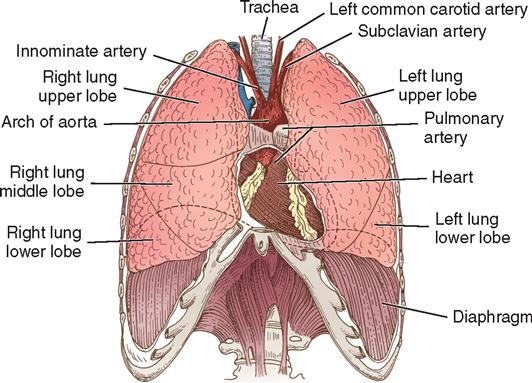
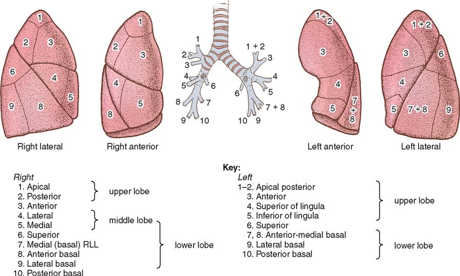
The lungs are the essential organs of respiration. The base of each lung rests on the diaphragm, whereas its apex (upper end) projects into the base of the neck at a level above the first rib. The bronchus, the nerves, the lymphatics, and the pulmonary and bronchial vessels enter and leave the lung on the mediastinal surface in a structure known as the hilum, or root, of the lung. Deep fissures divide the spongy, porous lung into lobes. The primary bronchi divide and then subdivide into each lobe and eventually become bronchioles. The right lung has an upper, a middle, and a lower lobe; the left lung has only an upper and a lower lobe (Figure 14-4). However, the lungs are similar in that each is composed of 10 major segments (Figure 14-5). Each segment extends to the pleural surface, expanding in volume from its center to its peripheral edges. Each segment also has its own bronchus and branches of the pulmonary artery and vein.
The bronchial arteries, arising from the aorta, supply nourishment to the lungs. They vary in their number and course. The arrangement may include two branches to the left lung and one branch to the right lung, which later branches into two, or there may be one or two branches for each lung. The pulmonary arteries carry the blood to the pulmonary parenchyma, and the pulmonary veins transport the oxygenated blood to the left atrium.
The nerves of the lungs are a part of the autonomic nervous system. They regulate constriction and relaxation of the bronchi and of the blood vessels within the lungs.
Although the thoracic cavity is an airtight space, the lungs receive outside air through the nasal passages, trachea, and bronchi. The main function of the lungs is to exchange carbon dioxide for oxygen. Normally, as the thorax expands, the lungs also expand as air is drawn in; during expiration, the thorax relaxes and the lungs passively contract as air is forced out. Inspiration normally takes place when the intrathoracic pressure is slightly below atmospheric pressure (76 cm Hg, or 760 mm Hg) and when a partial vacuum exists between the parietal and visceral pleural (intrathoracic) surfaces. As the muscles of inspiration contract to enlarge the chest cage, the lungs passively follow the diaphragm and chest wall because of decreased intrathoracic pressure. The acts of inspiration and expiration are the result of air moving in and out of the lung, causing pressure to equalize with that of the atmosphere at the end of expiration.
The normal intrapleural pressure varies from −9 to −12 cm H2O during inspiration and from about −3 to −6 cm H2O during expiration. The greatest amount of air that can be expired after a maximum inspiration is termed the vital capacity, and the volume of gas remaining in the lungs after maximal expiration is residual volume. Size, age, gender, and pulmonary disease of the patient influence vital capacity. Any condition that interferes with the normally negative intrapleural pressure affects respiratory function.
Surgical Technologist Considerations
The surgical technologist must be prepared for the demands of thoracic surgery. From bronchoscopy to mediastinoscopy and resection to lobectomy, surgical technologists’ responsibilities include prepping, draping, special equipment knowledge, instrumentation, suture, and supplies. It is important to note that thoracic surgery also usually entails special care and labeling of specimens, including frozen section, cytology, and cultures.
Medications on the sterile field may include a local injectable, as well as a warm antibiotic irrigation. Depending on the procedure, the surgeon and the anesthesia provider may decide to do a block or epidural for additional pain control. Many sizes and types of chest tubes will be needed, as well as stapling devices. There are many types of anastomotic devices on the market. It is important to familiarize yourself with what your facility has available for use by the surgeon.
Additional Considerations
During thoracic surgery, the surgical technologist is concerned with both preparatory patient considerations (e.g., verification of the patient, surgical procedure, and site; positioning; draping) and the requirements of the surgical intervention (e.g., medication delivery; instrument, equipment, and supply availability).
Positioning.
The type of position used in thoracic surgery is determined by the operative procedure planned. Bronchoscopy is performed in the supine position with a shoulder roll. Thoracotomy can be performed with the patient in one of three common positions: (1) lateral for the posterolateral approach, (2) semilateral for the anterolateral approach (Figure 14-6), and (3) supine for the median sternotomy approach. Arms are placed on padded armboards with the palms up and fingers extended. Armboards are maintained at less than a 90-degree angle to prevent brachial plexus stretch. If there are surgical reasons to tuck the arms at the side, the elbows are padded to protect the ulnar nerve, the palms face inward, and the wrist is maintained in a neutral position (Denholm, 2009). A drape secures the arms. It should be tucked snuggly under the patient, not under the mattress. This prevents the arm from shifting downward intraoperatively and resting against the OR bed rail.
Draping.
Draping may be minimal for bronchoscopic procedures. The principles of draping for other procedures are followed in thoracic procedures. Drapes may consist of a fenestrated sheet or single sheets surrounding the incision site. To prevent instruments from falling from the field, the surgical technologist may place a magnetic pad on the drapes below the incision site when the patient is placed in lateral position. A forced-air warming blanket is placed on the patient before draping to maintain normothermia. Sequential compression devices are often applied also to prevent the development of deep vein thrombosis (DVT).
Instrumentation.
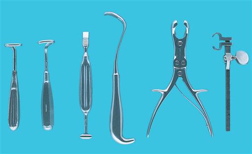
Bronchoscopy instruments are designed to directly inspect and observe the larynx, trachea, bronchi, or mediastinum; to remove secretions; to obtain washings or tissue for bacterial and cytologic studies; or to remove tissue. They are also designed to remove foreign bodies. Instrumentation for thoracic surgery includes the laparotomy instrument set (see Chapter 2) and specialty items. Instruments used for a thoracotomy or chest procedure include a combination of delicate and heavy instruments. Stapling equipment commonly is used as suturing devices and requires staplers and reload staples of appropriate sizes. The delicate instruments are used to cut tissue and vessels or to clamp tissue in an atraumatic manner. The heavier instruments are used for bone cutting, dissecting, or retracting. Instrumentation must also be available for hemostasis and suturing of all types of tissue.
The surgical technologist should determine the arrangement of items on the instrument table and Mayo stand; this arrangement should be an effective standard method that applies principles of work simplification and thorough knowledge of procedures. Lengthy incisions are often required for thoracic procedures; therefore it is critical that an instrument count be performed before closure.
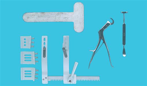
The back table will include instruments required for the type of thoracic surgery being performed. Common setup includes a basin set, thoracotomy/thoracoscopy set, and a self-retainer retractor, such as a Karlin or Beuford. Although surgeons may have a preference for the type of scope and camera used in endoscopic procedures, either monopolar or bipolar electrosurgery is commonly employed. Preferences will also include an aspirating needle, endo graspers, endo forceps, punches, or biting biopsy clamps, as well as minimally invasive needle holders and scissors. Extra long instruments should also be available.
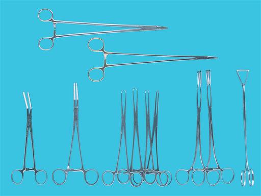
The Mayo stand includes long and standard Metzenbaum and Mayo scissors and long DeBakey, Russian, and Gerald forceps. Blades needed include 20, 10, 15, and 11 on both long and standard handles. Long hemoclip appliers in medium and large sizes, as well as an extended electrosurgical pencil (active electrode), and long silk ties on a pass are common. Clamps include Duvals, long Allises tonsils, and hemostats. The Allison lung retractor is the most common handheld thoracic retractor. Figures 14-7 through 14-9 show examples of instruments used in thoracic procedures.
A long double-armed monofilament suture is needed for any unexpected bleeding. Closing suture on an open procedure includes pericostal sutures. These are placed to appose the ribs and are commonly a braided absorbable suture material on a rather large needle.
Equipment.
In thoracic surgery, a variety of equipment is used, including a forced-air warming unit, fiberoptic headlights, fiberoptic light sources, video equipment, sequential compression devices, and anesthesia supplies. Double-lumen endotracheal tubes are commonly used for thoracotomies.
The neodymium:yttrium-aluminum-garnet (Nd:YAG) or CO2 laser can be used for treating tracheobronchial lesions with use of a bronchoscope. Obstruction of the mainstem bronchus and trachea caused by benign and malignant lesions can also be effectively treated with laser therapy. Use of laser equipment requires a thorough understanding of the equipment, the safety issues, the responsibilities, and the planned surgical procedure.
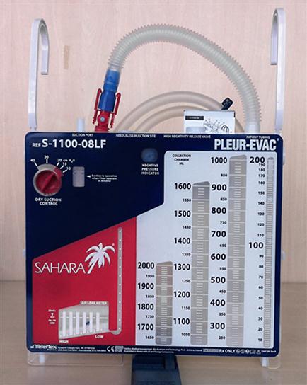
One or more chest catheters (tubes) may be inserted for postoperative closed-chest drainage. The chest tubes provide a conduit for drainage of air, blood, and other fluid from the intrapleural or mediastinal space and reestablishment of negative pressure in the intrapleural space. Drainage systems use three mechanisms to drain fluid and air from the pleural cavity: positive expiratory pressure, gravity, and suction. The chest tubes are connected to a sterile water-seal or gravity drainage system. Water-seal suction may be necessary when a persistent air leak cannot be controlled by drainage alone. Several compact, disposable units are available. The disposable units have three or four compartments for drainage, water seal, and suction. The first chamber collects the drainage from the intrapleural space, the second chamber provides the water seal, and the third provides the suction control determined by the level of water (Figure 14-10). If two chest tubes are inserted, they may be attached by a Y connector to a single drainage unit or may be attached individually to two separate units. Chest tubes are generally removed within 5 to 7 days.
Surgical Interventions
ENDOSCOPY (DIAGNOSTIC OR THERAPEUTIC)
Endoscopy refers to examination of hollow body organs or cavities with instruments that permit visual inspection of their contents and walls. The endoscopic procedures pertinent to thoracic surgery are bronchoscopy, mediastinoscopy, and thoracoscopy. Each endoscopist has preferences regarding the type of endoscope, positioning of the patient, type of anesthetic, and equipment. Invasive diagnostic or therapeutic measures enhance the decision to pursue surgical intervention by providing information related to the disease process, including histologic characteristics, location of the lesion, and lesion extent. Therapeutic endoscopy provides treatment by removal of the lesion or foreign body.
Standard Bronchoscopy Using Rigid Bronchoscope
Standard bronchoscopy is the direct visualization of the mucosa of the trachea, the main bronchi and their openings, and most of the segmental bronchi. It also includes removal of material for microscopic study if necessary.
Bronchoscopy is an integral part of the examination of patients with pulmonary symptoms such as persistent cough or wheezing, hemoptysis, obstruction, and abnormal roentgenographic changes. Common causes of bleeding (hemoptysis) are bronchiectasis, carcinoma, and tuberculosis. Congenital anomalies and suspected presence of a foreign body, especially in infants and children, are responsible for emergency examination of the respiratory tract.
The surgeon performs bronchoscopy to determine the presence of a lesion in the tracheobronchial passages, to identify and localize that lesion accurately, and to observe periodically the effects of therapy. It can be completed for dilating structures, debriding tumors, or evacuating clots. In suspected carcinoma, the aspirated secretions obtained by bronchoscopy may contain malignant cells.
Procedural Considerations.
Flexible bronchoscopy on an adult patient may be completed after induction of local (topical) anesthesia or monitored anesthesia care; a child usually receives a general anesthetic. Patients undergoing rigid bronchoscopy should be paralyzed and ventilation continued to minimize trauma. The adult patient administered a local anesthetic may experience discomfort and anxiety. To reduce anxiety, the perioperative nurse should introduce members of the surgical team, explain intraoperative activities, and provide reassurance to the patient. The oral structures, including the teeth and lips, should be assessed for integrity. Loose teeth may require removal before or during the procedure.
The anesthesia provider or nurse providing moderate sedation may administer IV sedatives or analgesics during the procedure. The topical (or local) anesthetic setup should include a headlight for visualization, laryngeal mirrors of various sizes, a lingual spatula, sprays with straight and curved cannulae, and anesthetic drugs, as ordered. Other items include the laryngeal syringe with straight and curved cannulae, Jackson cross-action forceps, and the Schindler pharyngeal anesthetizer, if desired. Luer-Lok 10-ml syringes and 20- and 22-gauge needles are needed for transtracheal injection. The surgical technologist ensures that all requested instrumentation is available to avoid delays and ensure the procedure is performed efficiently for the patient.
Anesthetic drugs frequently used for local or topical anesthesia are lidocaine (Xylocaine), procaine (Novocain), and tetracaine (Pontocaine, Cetacaine) with or without epinephrine. Pauses of 3 to 4 minutes are taken between applications of the anesthetic agent to the tongue, palate, and pharynx and then to the larynx and to the trachea. The surgeon or anesthesia provider applies the anesthetic agent by means of a spray or laryngeal syringe with a straight or curved cannula.
Some physicians prefer to have the patient sit upright and gargle with the topical anesthetic mixture, rinse it around in the mouth, and then expectorate it, thereby producing a partial anesthesia of the buccal mucosa and pharynx.
For direct bronchoscopy, the surgeon or anesthesia provider uses a long metal cannula attached to a syringe to apply the anesthetic agent to the surface of the vocal cords; then the agent is injected through the anesthetized glottis into the trachea. This act causes the patient to produce a sharp, sudden cough. For intrabronchial anesthesia, a portion of the anesthetic agent is introduced through the bronchoscope.
The patient may be positioned either in the supine position—with the shoulders elevated on a small roll, gel pad, or a sandbag to gently extend the head and neck—or in the sitting position.
The setup includes the bronchoscope, telescopes of desired types, fiberoptic light cords, and the fiberoptic light source. Each standard scope requires a fiberoptic light carrier, cord, and light source. The surgical technologist ensures that duplicates of each, along with the appropriate replacement lightbulbs for the light source, are available for immediate use. The light source should be tested periodically and immediately before use. To test the fiberoptic light carrier and telescope, the instrument should be held vertically by the ocular end. The endoscope should always be tested immediately before passage into the patient. The light-intensity dial should be set at the proper level, as specified by the manufacturer. The light source should be switched on and off to test its function. During a procedure, the perioperative nurse switches the light source to standby mode whenever it is not in active use to conserve the life of the bulb and promote cooling.
Other supplies that will be needed are suction tubing, aspirating tubes, specimen collectors, sponge carriers, and the desired type of forceps. The metal sponge carrier consists of two parts: an inner rod, which has two jaws protruding from its distal end; and an outer band, which is screwed down on the inner rod so that a sponge can be held securely within the jaws. Small gauze sponges are used to keep the field dry, remove secretions, and apply a topical anesthetic agent. Cytologic specimen collectors, such as the Clerf or Lukens, are used to hold secretions as they are obtained. Aspirating tubes of different lengths and designs are used to remove secretions and collect material for microscopic examination and culture(s). The straight aspirating tube with one or two openings at the distal end is used to remove material from the pharynx, larynx, and esophagus. The curved aspirating tube with a flexible tip is used to remove secretions from the upper and dorsal orifices of the bronchi.
The surgeon uses various types of forceps to remove foreign bodies or tissues for histologic study. Biting-tip forceps may be used to secure tissue for pathologic study. Forceps with jaws that veer laterally at about a 45-degree angle from the instrument’s axis permit visualization during the biopsy maneuver. Bronchoesophageal forceps consist of a stylet, a cannula with a handle, a screw, a locknut, and a setscrew. Forceps for laryngeal and bronchial regions are designed to remove tissue specimens. The circulating nurse will prepare several specimen containers with the identifying patient information label, along with laboratory slips for specific requests. If brush specimens are collected, slides and alcohol are required. Specimen safety practices are followed, and the surgical technologist and circulating nurse should “repeat back” the type and location of each specimen collected.
The standard bronchoscope is a rigid speculum used for visualizing the tracheobronchial tree. The rigid bronchoscope might be selected for biopsy of a large central mass, for removal of a foreign object, or to control hemorrhage during biopsy of a vascular mass. The rigid bronchoscope remains the instrument of choice for removal of foreign bodies in infants and children. A fiberoptic light carrier is inserted into the bronchoscope to illuminate the distal opening. A side channel is incorporated into the bronchoscope to permit aeration of the lungs with oxygen or anesthetic gases. An additional device, the Sanders Venturi system, which is available to the anesthesia provider, provides adequate patient ventilation during bronchoscopies and laryngoscopies.
The endoscopist risks contamination in the presence of communicable diseases. For this reason, the endoscopist and assistants should wear facemasks and eye protection or wear a transparent shield attached to a headband. With increasing numbers of patients with tuberculosis, particulate respirators are recommended as protective devices. Aseptic technique is used to prevent cross-contamination.
Operative Procedure
Stay updated, free articles. Join our Telegram channel

Full access? Get Clinical Tree


