CASE 14
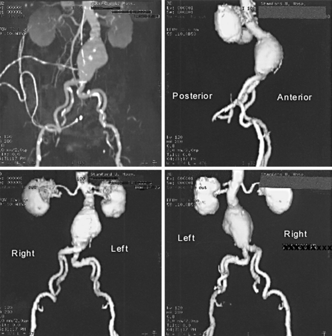
A 49-year-old man presented to his physician complaining of lower back pain. The pain has lasted for 3 days and is unaffected by movement, though the pain is alleviated somewhat when the patient’s thighs are flexed. When the anterior abdominal wall was palpated, a tender pulsatile mass was detected just superior to the umbilicus. The physician also detected diminished pulses in the femoral and dorsalis pedis arteries, and bruits were heard over the palpable mass. Imaging studies showed an abdominal aortic aneurysm (Fig. 3-1) with atherosclerotic lesions of the common iliac arteries. The aneurysm was surgically repaired by the insertion of a synthetic prosthesis.
ANATOMICALLY, WHERE DO AORTIC ANEURYSMS COMMONLY OCCUR?
Most aortic aneurysms occur below the level of the renal arteries (Figs. 3-1 and 3-2). Less frequently the aneurysm involves the suprarenal abdominal aorta and thoracic aorta.
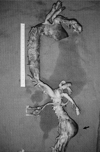
FIGURE 3-2 Human aorta showing an infrarenal abdominal aortic aneurysm at the arrow.
(Towsend C et al.: Sabiston Textbook of Surgery, 17e. WB Saunders, 2004. Fig. 64-2.)


