13. Plastic and Reconstructive Surgery


Plastic and Reconstructive Surgery  


LEARNING OBJECTIVES


After studying this chapter the reader will be able to:


Overview


Derived from the Greek word plastikos, which means to mold or give form, plastic surgery is a medical specialty that restores or gives shape to the body. There are two different subspecialties of plastic surgery. Cosmetic surgery restores or reshapes normal structures of the body, to improve appearance and self-esteem. Reconstructive surgery treats abnormal structures of the body caused by birth defects, developmental problems, disease, tumors, infection, or injury to restore function and correct disfigurement or scarring (American Society of Plastic Surgeons [ASPS], 2009a.) As a surgical specialty, plastic surgery owes much of its heritage to knowledge gained from the wars of the twentieth century (History box).


Despite the economic downturn, 12.1 million cosmetic surgical procedures were performed [by surgeons certified by the American Society of Plastic Surgeons (ASPS)] in 2008, an increase of 3%; the majority were minimally invasive procedures. Females constituted 91% of all patients undergoing cosmetic procedures. Hispanic, Asian, and African American patients showed an increase in cosmetic procedures; the number of Caucasians undergoing cosmetic procedures decreased by 2%. Office-based cosmetic procedures increased by 13%, with a total of $10.3 billion dollars spent on the cosmetic procedures in the United States (Ambulatory Surgery Considerations). The 4.9 million reconstructive procedures also showed a 3% increase, although breast reductions decreased by 16% (ASPS Stats, 2009).


Surgical Anatomy


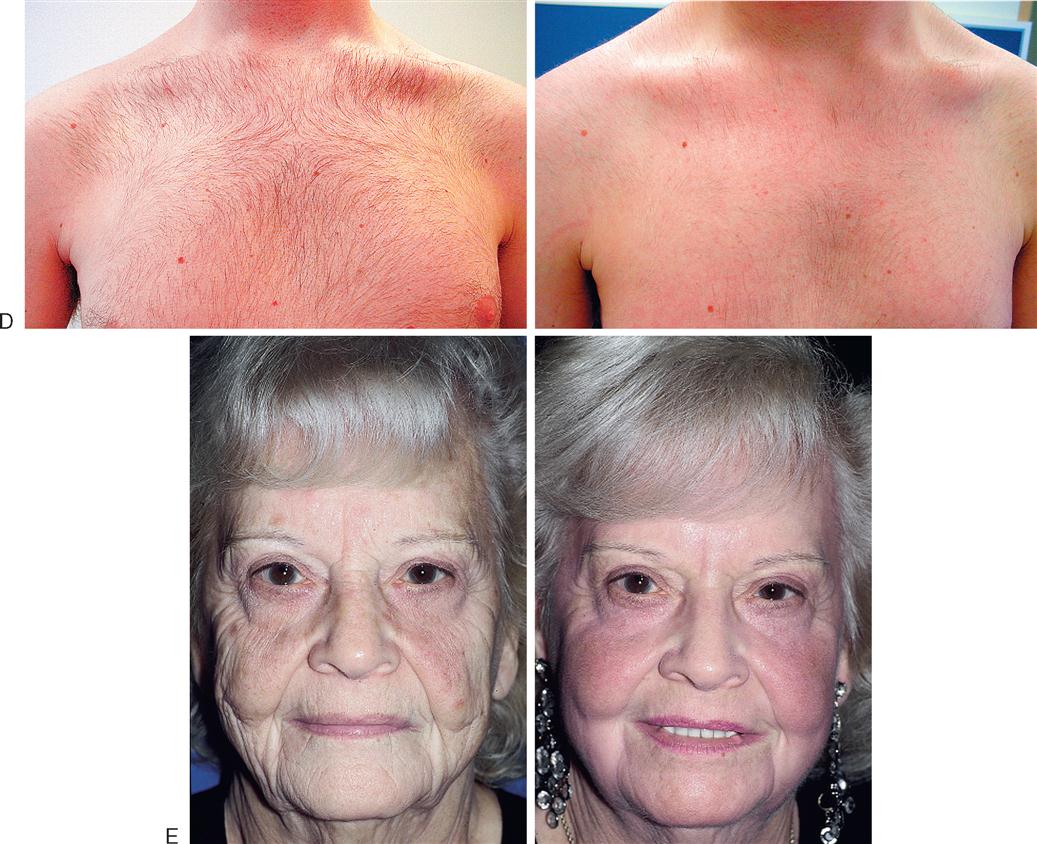
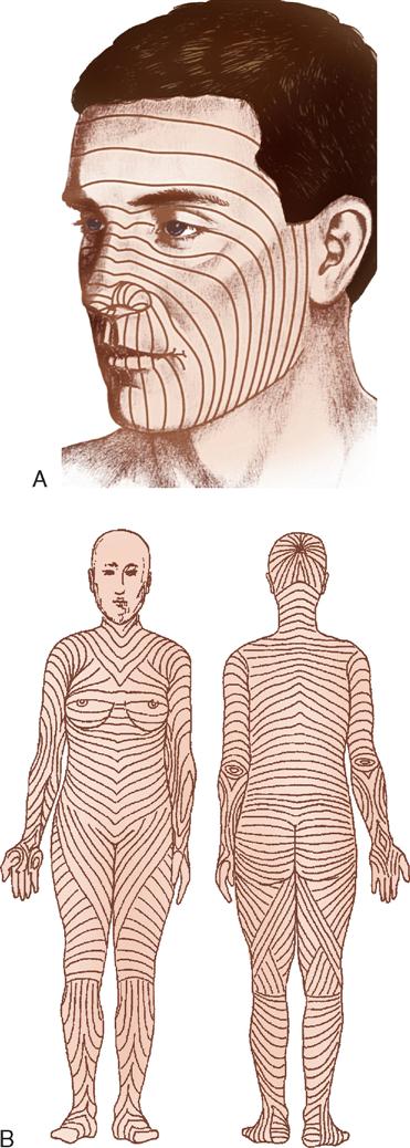
Plastic and reconstructive surgery is not limited to a single anatomic or biologic system. It is based on thorough understanding of the anatomy and biology of tissue. Operative techniques are complex and staged to achieve the expected results. The surgery also involves removing, reducing, enlarging, and recontouring, as well as camouflaging scars into existing skin lines (Figure 13-1). The tissues of the body can be transferred to use as various types of flaps. Free flaps are the transfer of tissue along with its vascular pedicle. When nerve is anastomosed with these flaps, they are called neurovascular free flaps. Flaps are used to cover defects or create new structures such as breasts, digits, or facial structures. Body parts can also be transplanted. By improving the patient’s deformity, the patient’s self-esteem will improve and the patient will feel more comfortable in public and social activities. The body changes as we age. The patient’s concern with aesthetics, the variety of acquired defects, the diversity of operative techniques, and the psychologic responses of patients offer unique learning experiences and challenges for perioperative patient care.


Surgical Technologist Considerations


Preparation of the OR Suite.


Assemble all necessary medical and surgical supplies, equipment, suture material, positioning aids, implantable devices, and medications. Ensure that lights and video equipment are in working order, that emergency supplies are present, and that compressed gases are adequate. Depending on the procedure to be performed, the OR bed may need to be configured differently from the standard room setup. Plastic and reconstructive surgeons frequently use preoperative photographs of the patient when attempting to restore or modify appearance. These photographs help the surgeon maintain perspective since features may change because of surgical positioning. Preoperative and postoperative photos, in order to be accurate, should be taken with the same lighting, angle, and distance (Hagan, 2008).


Equipment and Special Mechanical Devices.


Essential equipment for any OR includes a fully functional bed that may be positioned for any number of special needs and also has accessory attachments, such as headrests and aids for extremity positioning. The room must also have well-positioned and numerous electrical outlets, good overhead lighting, suction equipment, mounted x-ray view boxes, and computer terminals for those facilities using electronic medical records. Step stools, tables, chairs, hand tables, tourniquets, microscopes, and intravenous (IV) poles should be in appropriate supply and accessible. Surgeons often provide their own digital cameras although facilities may have them available for use.


INSTRUMENTATION.

Basic instrument trays are available for the plastic surgery OR. A “local” tray may include Bishop Harmon and adson tissue forceps (with and without teeth); straight and curved iris, Stevens, and Metzenbaum scissors; fine mosquito forceps; and skin hooks. Minor and major trays for plastic surgery may contain a range of tissue forceps, scissors, hemostats, and retractors. With the addition of instruments for specific surgeries, these trays usually suffice for all plastic surgery operations. Adequate instrumentation should be available to avoid flash sterilization (Risk Reduction strategies).


HISTORY


Origins and Growth of Plastic Surgery


Historically, battlefield combat has been an impetus for the development of new medical and surgical techniques based on the injuries sustained. As battlefield technology and the weapons of war became more sophisticated throughout history, the degree of injury and tissue devastation became more horrific. The trench warfare of World Wars I and II gave rise to a whole new category of facial injuries. Helmets protected combatant’s skulls and the trenches offered some protection to the chest, but the face was exposed, and as a result, devastating burns and fractures of the face occurred. Special hospitals were created to address these problems, and even before the United States joined the first World War, the Harvard unit sent 35 physicians and surgeons, 3 dentists, and 75 nurses from various medical centers to assist in caring for the wounded. These visionaries were soon developing new techniques and procedures to correct the disfiguring injuries. They were credited as being the first generation of modern plastic surgeons, and helped give much needed respect to the specialty.


While World War I helped reinvent plastic surgery, the specialty has been identified as long ago as 600 BC, when a Hindu surgeon described using a cheek flap to reconstruct a nose. Another flap technique, this time using the forehead to reconstruct a severed nose, was performed around 1000 AD in India. The Italian surgeon Gaspare Tagliacozzi, also known as the “father of plastic surgery,” developed still another flap surgery using the upper arm to reconstruct a nose. History tells us that the condition of the nose, whether from war, punishment, or social disease (syphilis), presented a story that often was undesirable. This resulted in impetus in the different societies to camouflage the injuries, thus propelling advances in plastic surgery.


Following the end of the first World War, plastic surgery turned its attention to the rest of society, and concentrated on deformities caused by birth or trauma. Soon, some surgeons began using their talents to improve less than desirable facial features. For example, Fanny Brice underwent a rhinoplasty in her apartment in 1923 to change the appearance of her nose from “prominent to decorative.” In 1924 a New York newspaper had a contest to transform the city’s homeliest woman into a beauty. Dr. John Howard Crum performed the first facelift on record in the Grand Ballroom of the Pennsylvania Hotel in New York City in 1931, during which “a pianist accompanied him with appropriate popular tunes, flashbulbs popped, and men and women fainted.”


Modified from Feldman E: Before & after: cosmetic surgery was born 2,500 years ago and came of age in the inferno of the western front, Am Herit 55(1):60-70, 2004; The history of plastic surgery: the early years, available at www.plasticsurgery.org/About_ASPS/History_of_Plastic_Surgery.html. Accessed July 6, 2009.


AMBULATORY SURGERY CONSIDERATIONS


Patient Choices in Facility


There has been a dramatic shift toward encouraging patients to be more involved and informed in all of their healthcare decisions. Many organizations are focusing on active participation of patients in their own care. One aspect of this is choosing the outpatient facility for their plastic surgery. In keeping with this trend, the American Society of Plastic Surgeons (ASPS) has developed a brochure, with an excerpt available on-line, to help patients choose a facility. The brochure includes the following information:


♦ Types of facilities: For ambulatory (discharge same day as surgery) procedures, the facility may be part of the surgeon’s office, a surgical suite adjacent to the office, a free-standing facility, or part of a hospital.


♦ Choice and benefits: Some patients prefer no overnight stay at a hospital. Cost savings and convenience along with added privacy and personalized care may all be factors.


♦ Accreditation: As a result of the strict guidelines for equipment, staff, anesthesia services, and hospital access, patients are encouraged to determine if a facility is accredited, as it could indicate the quality of the facility. (All ASPS member surgeons are required to operate in an accredited medical facility.)


♦ Insurance: The accreditation mentioned above also impacts financial aspects. Although facility fees are typically not covered by insurance providers for elective procedures, facility fees for reconstructive procedures may be covered. Reimbursement may be expedited for facilities that carry accreditation because the accreditation ensures that certain quality standards are met at that facility. Also, similar services at a hospital generally cost more than in an ambulatory facility.


♦ Individual considerations: Each patient must discuss the options with his/her surgeon. Depending on medical history and condition requiring surgery, ambulatory surgery may not be appropriate for everyone.


Modified from American Society of Plastic Surgeons: Ambulatory facilities: same day facilities: choosing same day surgery facilities, Arlington Heights, Ill, 2008, available at www.plasticsurgery.org/Patients_and_Consumers/Planning_Your_Surgery/Same_Day_Surgery_Facilities.html. Accessed June 17, 2009.


DERMATOMES.

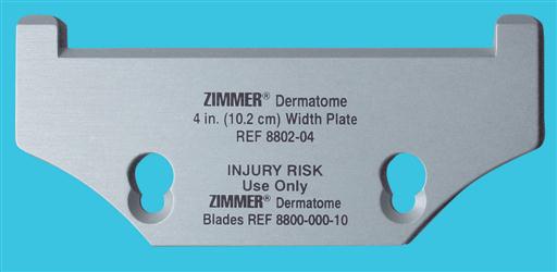
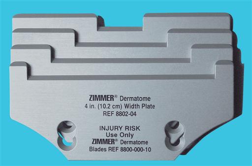
Dermatomes are used for removing split-thickness skin grafts (STSGS) from donor sites; they are of three basic types: knife, drum, and motor-driven (Figures 13-2 through 13-5). Sterile mineral oil and a tongue blade should be available when stsgs are being obtained.



SKIN MESHERS.

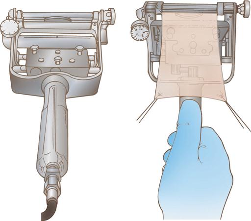
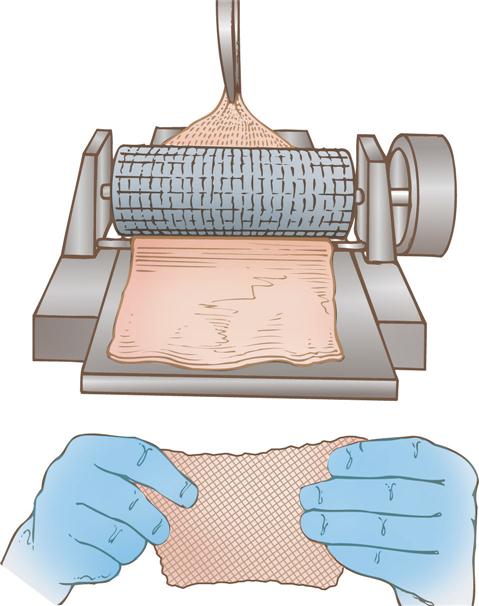
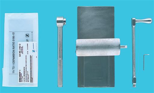
Several types of skin meshers are available. Each is designed to produce multiple uniform slits in a skin graft, approximately 0.05 inch apart. These multiple apertures in the graft can then expand, permitting the skin graft to stretch and cover a larger area. Meshing also facilitates drainage through the graft, preventing fluid accumulation under a graft. The graft is placed on the carrier and passed through the mesher (Figures 13-6 and 13-7). The manufacturer supplies sterile carriers for meshers. They are usually available in several sizes, which determine the expansion ratio of the skin graft.


PNEUMATIC-POWERED INSTRUMENTS.

Pneumatic-powered instruments use an inert, nonflammable, and explosion-free compressed gas as their power source. The motor may be activated by a foot pedal or hand control. The various attachments should be sterilized as recommended by the manufacturer to prolong instrument life and ensure effective sterilization. The following attachments may be used in plastic surgery:


A pneumatic tourniquet with an inflatable cuff is used in most hand surgery procedures as well as in other upper and lower extremity surgical interventions. The tourniquet is described in Chapter 10.


ELECTROSURGICAL UNIT.

The electrosurgical unit (ESU) is employed and safety precautions should be followed. Monopolar ESU will require the use of a grounding pad (return electrode/dispersive electrode). Specialized tips will be used with the ESU pencil including needle point and guarded along with the standard tip. The use of specialized tips allows the use of higher coagulation settings to obtain hemostasis with little or no effect on adjacent tissue or structures. Inspect ESU tip and cord on handpiece for damage before use, be sure tip is securely seated into the place handpiece in nonconductive holster when not in use, keep tip free of eschar or tissue build-up. Methods to do so include moistened sponge, instrument wipe, and abrasive electrode cleaner, but not a scalpel blade.


BIPOLAR COAGULATION UNIT.

Bipolar electrosurgery is the use of electrical current in which the circuit is completed by means of two parallel poles located close to one another. One pole is positive; the other is negative. The flow of current is restricted between these two poles, which are usually the tines of the bipolar forceps. Because the poles are so close, low voltages are used to achieve the tissue effect. Because electrical current does not flow through the patient, a return electrode (dispersive pad) is not necessary. This makes bipolar electrosurgery very safe and permits precise electrocoagulation.


LASERS.

A variety of lasers are employed for plastic surgical procedures. The perioperative team must ensure that the laser safety accessories specific to the type of laser being used are available. Types of lasers and their common uses are presented in Box 13-3, pp. 637–639.


BOX 13-3


Lasers in Plastic Surgery


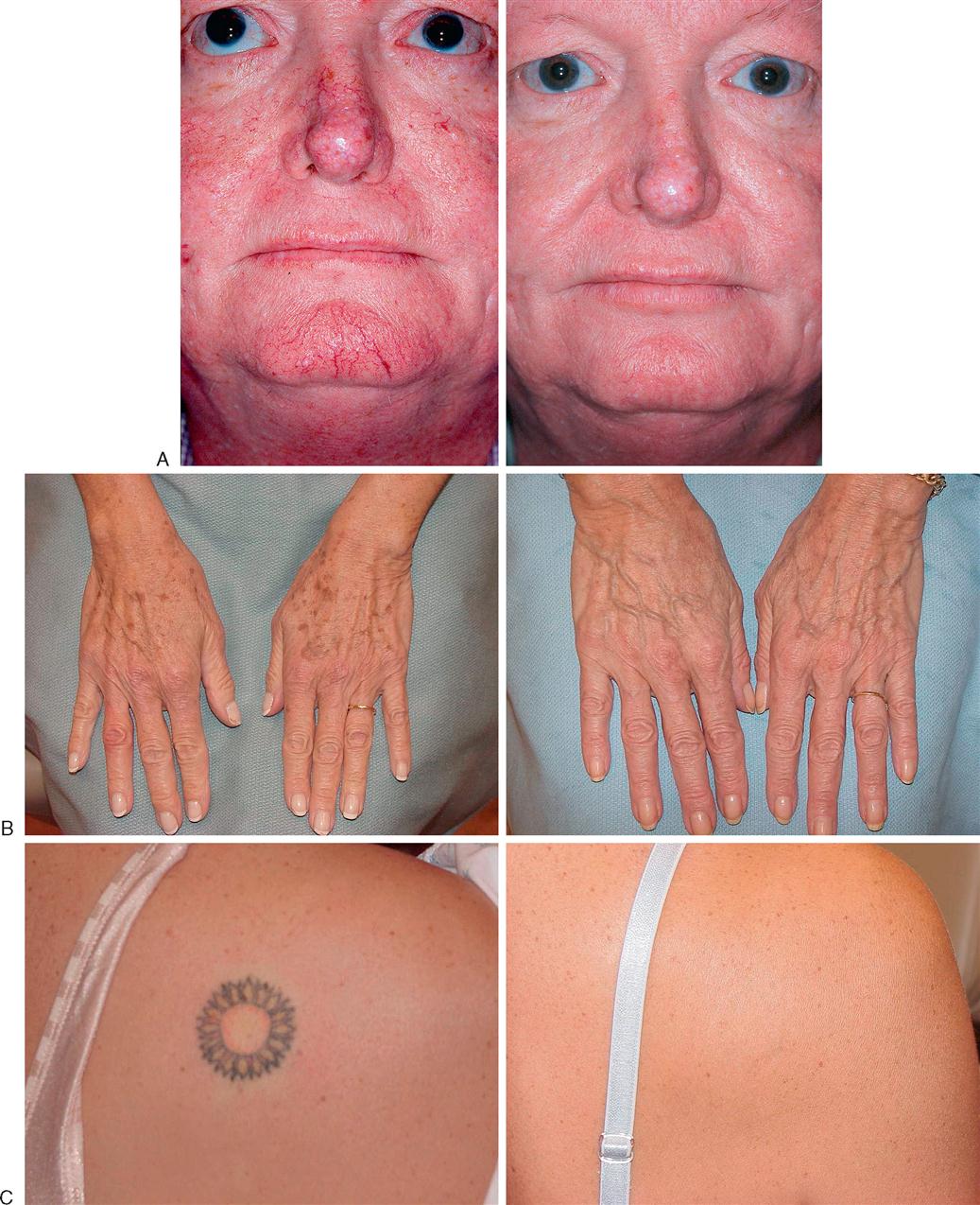
Common uses for lasers in plastic surgery include exfoliation, treatment of vascular malformations, removal of hair and tattoos, and tightening of collagen fibers in aging skin. A variety of lasers are available; selection of the appropriate laser is dependent on the patient’s diagnosis because the effect of the laser on the skin tissue is dependent on its wavelength. The types of lasers and their common uses are described below.


Laser Use
CO2 Desiccation of benign lesions of skin, skin resurfacing, cutting tissue
Excimer Eye surgery, psoriasis
Argon Hemangiomas, telangiectasias
Nd:YAG Benign pigmented lesions and red tattoos
Candela dye Tattoos, pigmented benign lesions, hemangiomas
Helium-neon Biostimulation, wound healing alleviation, acupuncture
Diode Hair removal, tattoo removal
Ruby Nevi removal, dark tattoos
Erbium: YAG Rhytides
Q-Switch Benign pigmented lesions and dark tattoos


Modified from Choi JE et al: Treatment of Becker’s nevi with a long-pulse alexandrite laser, Dermatol Surg, April 27 [Epub ahead of print], 2009; Kono T et al: Long-pulsed neodymium:yttrium-aluminum-garnet laser treatment for hypertrophic port-wine stains on the lips, J Cosmet Laser Ther 11(1):11-13, 2009; Krupashankar DS: Standard guidelines of care: CO2 laser for removal of benign skin lesions and resurfacing, IADVL Dermatosurgery Task Force, Indian J Dermatol Venereol Leprol 74(suppl):S61-S67, 2008; Mendonca DA et al: Venous malformations of the limbs: the Birmingham experience, comparisons and classification in children, J Plast Reconstr Aesthet Surg, Dec 27 [Epub ahead of print], 2008; Onesti MG: Surgical and laser treatment of Sturge-Weber syndrome, Aesthet Plast Surg, Mar 19 [Epub ahead of print], 2009.


FIBEROPTIC INSTRUMENTS.

Examples of fiberoptic instrument attachments used in plastic surgery are a headlight for rhinoplasties, augmentation mammoplasties, and other procedures; a mammary retractor for augmentation mammoplasties; a rhytidectomy retractor; abdominoplasty retractors; and endoscopic face and forehead fiberoptic instrumentation.


LOUPES.

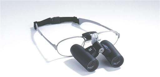
Loupes (Figure 13-8) are magnifying lenses used by many plastic surgeons for microvascular surgery and nerve repairs and for numerous other instances in which cosmetic results are improved by the magnification effect. The nurse should inquire about the use of loupes before the surgeon dons a headlight because adjustments will need to be made to the headlight alignment if the loupes are required in midprocedure. Adjusting or removing the headlight in midprocedure has the potential to contaminate the sterile field.


MICROSCOPE.

The microscope is frequently used in nerve repairs and microsurgical anastomoses; the nerves or vessels to be repaired, such as in hand surgery, and the suture used to do so (sometimes 9-0, 10-0, or even 11-0 size) can be finer than human hair and thus requires magnification. While each microscope has different features, an important matter to avoid confusion is whether the surgeon control overrides the assistant view, or if each can separately adjust the field of view.


WOOD’S LAMP.

The wood’s lamp is an ultraviolet lamp used in a darkened room to determine the viability of skin flaps. After IV injection of fluorescein, the blood vessels appear bright purple (the skin appears yellow). Sodium fluorescein is excreted in the urine, and patients should be informed of this.


SPECIAL SUPPLIES.

Surgeon-specific and procedure-specific special supplies are frequently added to instrument setups for plastic and reconstructive procedures. these commonly include the following: sterile marking pen or methylene blue; ruler; local anesthetic of choice for injection, with syringes and needles; and ESU, with active electrode (pencil) and tip of choice, with tip cleaner.


Sutures.


Sutures range from permanent to absorbable and include monofilament and multifilament materials. The surgical technologist should be a good steward of costly resources and should verify the type and number of sutures needed before opening suture packages, as well as needle preference, to prevent waste. Many plastic surgical procedures have multiple techniques, each of which necessitates very specific suture choices.


Dressings.


Dressings are an essential part of the operative procedure in plastic surgery and may contribute to the ultimate outcome of the surgical intervention. Dressings are usually applied while the patient is still anesthetized. In general, the dressing should accomplish the following five goals:


Pressure dressings may be used to eliminate dead space, to prevent seroma and hematoma formation, and to prevent third spacing associated with liposuction and reconstructive procedures involving transfer of large muscle or tissue flaps. In some cases pressure can be achieved by the use of catheters or drains placed within the operative site and connected to closed-wound suction devices, such as a Hemovac or Jackson-Pratt. In smaller wounds a butterfly cannula may be inserted into the operative site, with the needle end placed into a red-top tube, such as a blood collection tube, that has a vacuum (evacuated tube).


Common general dressings and supplies available in sterile form and various sizes:


♦ Nonadherent gauze (e.g., Betadine gauze, Adaptic, Nu Gauze, Xeroform, Biobrane, Scarlet Red)


♦ Petrolatum gauze, ½ inch (or other packing material, such as Merocel sponge for nasal packing)


♦ Telfa


♦ Fine mesh gauze


♦ Interface


♦ Gauze dressing sponges, 4 × 4 inches, 2 × 2 inches


♦ Kling, Kerlix fluff, and Kerlix gauze rolls (2, 4, and 6 inches wide)


♦ Abdominal pads (most commonly used are 5 × 8 inches)


♦ Cotton sheets and balls


♦ Webril


♦ Tape (paper; silk; and foam; skin tapes, flesh-colored and regular [1/8, 1/4, 1/2, and 1 inch wide])


♦ Benzoin spray or swab or Mastoplast


♦ Ace bandages


♦ Coban


♦ Casting supplies and splints (as required for postoperative immobilization)


♦ Abdominal binders and other postoperative garments


♦ Slings


In some instances, such as a free flap, transparent dressings are used so that the flap can be monitored and observed for vascular flow. Compression garments and support devices are also frequently used by plastic surgeons. Proper fit is essential to minimize vascular compromise. Compression garments are typically applied over a light dressing. A proper garment is selected based on its characteristics (e.g., fabric, stretch, softness, antimicrobial properties) and proper sizing according to measurement instructions. Educating patients of the needs and benefits of compression garment use as well as providing hints for their proper application (avoid ripping with long nails, instructions on how to don the garment) promotes comfort and compliance (Gladfelter, 2007).


Implant Materials.


The range of materials available for implantation and augmentation in the specialty of plastic and reconstructive surgery has benefited from ongoing research. The perioperative team is responsible for complying with tracking regulations for implantable materials and devices (Patient Safety).


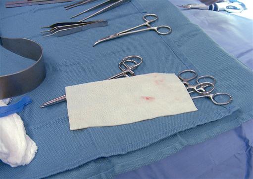
Biologic materials (autogenous grafts) are preferred when available. Autologous human tissue successfully utilized includes fat, solid dermis, and collagen. Human cadavers are used as a source for acellular collagen (AlloDerm) (Figure 13-9). This product is available in various sizes of sheeting and must be rehydrated in several steps. AlloDerm integrates with the body’s tissue and helps to prevent rejection over the long term.

image
FIGURE 13-9 AlloDerm.

Implant failure may be directly linked to bacterial contamination; therefore meticulous aseptic technique with minimal handling is essential when using implants of any sort. Most alloplastic implants are presterilized from the manufacturer.


Anesthesia.


A variety of anesthesia techniques are employed with plastic surgery procedures. Local, regional, tumescent, conscious sedation, deep sedation, and general anesthesia may be used, depending on the type of procedure, the patient’s anesthetic history, the American Society of Anesthesiologists (ASA) physical status classification, and the surgeon’s preference.


PATIENT SAFETY


Tracking Medical Devices


A variety of implantable devices are used in aesthetic and reconstructive plastic surgery procedures. Tracking of these devices is critical to patient safety. The manufacturer of the device must have a mechanism to locate implantables after they have been distributed. Devices may be recalled for sterility issues, malfunction, or any event that is found to pose a serious health risk.


The U.S. Food and Drug Administration (FDA) regulates the process of tracking medical devices and directs the tracking of devices whose failure would result in serious, adverse health consequences; devices that are intended to be implanted in the human body for more than 1 year; and devices that are life-sustaining and life-supporting and are used outside of a facility such as a hospital, nursing home, or ambulatory surgery center.


The perioperative team plays an important role in the accurate documentation of implantable devices for tracking purposes. Information that is typically gathered for tracking purposes includes:


If an implantable device is sterilized within the sterile processing department of the facility, monitoring requirements include the use of a process challenge device containing a biologic indicator. The load should be quarantined until the result of the biologic indicator is determined. Documentation should include a record of the sterilizer load identification number on the patient’s medical record, or the patient’s name on the load record. Lot identification provides a method for tracing problems in the event of a recall. Flash sterilization of implantable devices is not recommended.


Patients have the right to refuse tracking of their devices and may refuse to have their social security number used for tracking. The patient’s consent for tracking should be obtained before the procedure. If the patient refuses to have the device tracked, the nurse will document the refusal along with the required product information and report these data to the manufacturer.


Under the Safe Medical Device Act, institutions must also report any incident of death or serious injury relating to the use of a medical device. The FDA has classified and identified more than 1700 different devices that must be reported if the device is suspected of causing serious injury or death to an individual. Surgical technologists and nurses should work within their institutional policies to report these incidents.


Modified from Association for the Advancement of Medical Instrumentation: Comprehensive guide to steam sterilization and sterility assurance in health care facilities, Arlington, Va, 2006, AAMI; Chard R: Clinical issues, AORN J 90(1):117, 2009; Denholm B, Downing D: Clinical issues, AORN J 87(2):432-434, 2008; U.S. Food and Drug Administration: Medical device tracking, guidance for industry and FDA staff, available at www.fda.gov/cdrh/comp/guidance/169.html. Accessed July 6, 2009; U.S. Food and Drug Administration: Medical device reporting, available at www.fda.gov/cdrh/devadvice/351.html. Accessed July 6, 2009.


Preoperative Skin Preparation.


Most surgical interventions require that the operative site and adjacent areas be cleansed before surgery. The physician may prescribe that the patient carry out this treatment before surgery. Special attention is given to the fingernails for patients undergoing hand surgery; to hair for surgery of the head, face, or neck; and to oral hygiene for surgery in or near the mouth. Shaving is avoided and clippers, not a razor, are used if needed, because shaving creates an access for the entry of bacteria into the operative site (Health Stream, 2007). The eyebrows and eyelashes, in particular, are left intact to preserve facial appearance and expression. The surgical site is marked before surgery by the surgeon to designate the correct site and to define landmark areas. Either a povidone-iodine solution, an iodine-alcohol mixture, chlorhexidine gluconate (CHG), or another broad-spectrum agent may be selected for the antimicrobial skin prep. The use of CHG should be avoided around the ears and eyes. It is important to place shields on the eyes if prepping the periorbital site or performing an extensive head and neck prep, place plugs in the ear canals, and prevent pooling of the prep agent. When prepping for a skin graft procedure, separate skin prep setups are needed for the graft and donor sites.


Positioning and Draping.


The OR bed must be positioned so that the remaining space in the room can comfortably accommodate anesthetic equipment, members of the surgical team, instrument tables, and any adjunct equipment (hand table, drills, microscope, laser) to be used. The patient is carefully positioned on the OR bed so that all operative sites may be appropriately exposed and the airway easily observed and accessed.
SURGICAL TECHNOLOGY PREFERENCE CARD


Plastic and reconstructive surgery patients will range from infant to adult and include corrections for birth defects, trauma, burns, cancers, and cosmetic reasons.


Planning with the perioperative team is essential in plastic and reconstructive surgery with a wide variety of surgical procedures and patient populations. Specialty supplies, instruments, and prostheses will be used, frequently requiring strict sterile technique and efficient room layout. Allowing additional time preoperatively to organize the room and check the availability of supplies and prostheses provide for efficient communication between the circulator and scrub during the procedure. Additional setups for some procedures may be needed, so additional furniture and equipment should be organized to ensure sterility and no mixing of instruments.


Room Prep: Basic operating room furniture in place, thermoregulatory devices, padding, positioning supplies, ESU, and in some cases laser or a microscope


Prep Solution: In room and may require more than one site prepped


Catheter: In the room and correct size for patient


PROCEDURE CHECKLIST


Instruments


Specialty Suture


Specialty Supplies


Additional Supplies: both sterile and nonsterile


Medications and Irrigation Solutions


Drains and Dressings


Specimen Care


Before opening for the procedure, the surgical technologist should:


When opening sterile supplies:


Correct draping procedures depend on the location of the operative site or sites. Disposable drapes are often used because of their barrier qualities, ease of handling and storage, and versatility in adapting to a variety of plastic surgery procedures. Two of the most frequently used draping techniques in plastic surgery are the head drape and the hand drape. Both of these draping techniques have the goal of providing maximum mobility of the operative part. The head drape includes a fluid-resistant drape that encircles the head and the addition of a drape to cover the remainder of the body. The following techniques represent methods of obtaining maximum accessibility and sterile coverage for facial surgery:


1. A barrier sheet, folded in half, and two towels are placed beneath the patient’s head with the towels uppermost. The folded barrier sheet covers the headrest or head portion of the OR bed. One towel is brought around the patient’s head on each side to cover all hair, leaving the entire face (and ears, as necessary) exposed; the towel is then secured with nonpenetrating towel clamps. For craniofacial procedures a towel folded lengthwise in quarters may be placed under the head to assist with moving the head from side to side. Two additional towels are then placed diagonally across the neck, just under the chin; they are secured to each other (with nonpenetrating towel clamps) in the middle over the neck and are secured on each side to the towel around the head. A full sheet is then added to cover the patient from neck to feet.


2. After the head portion of the drape is placed, a split, or U, drape is added to cover the patient from neck to feet.


Surgical Interventions


Reconstructive Plastic Surgery


Reconstructive plastic surgery seeks to restore or improve function after trauma, disease, infection, congenital anomalies, or acquired defects while trying to approximate an aesthetic appearance.


REMOVAL OF SKIN CANCERS


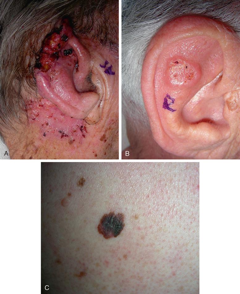
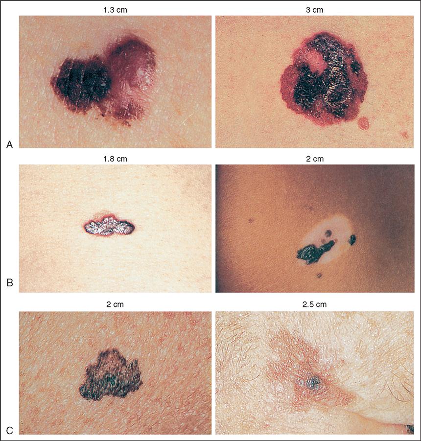
The estimated number of new skin cancer cases diagnosed in 2009 is 1 million (National Cancer Institute [NCI], 2009). The three most common skin cancers are basal cell, squamous cell, and melanoma (Gutierrez and Peterson, 2007). Basal cells account for approximately 70% of all skin cancers (Figure 13-10, A). If basal cell cancer is left untreated, it will grow locally, but rarely metastasizes (Box 13-1). Treated early, it may be cured by simple excision and closure (with pathologic diagnosis to ensure disease-free margins). Squamous cell skin cancers are considered more aggressive (Figure 13-10, B). Surgical treatment is the same as that for basal cell carcinomas. Melanoma accounts for the smallest percentage of skin cancers (5%), but it is treated much more aggressively because of its high mortality rate, comprising 75% of skin cancer deaths (Sladden et al, 2009) (Figure 13-10, C). Excision of melanoma may involve sentinel node mapping and excision. Early diagnosis of melanoma is imperative to successful treatment (Evidence for Practice). A Cochrane Database Systematic Review is underway (Sladden et al, 2009) since various international organizations varied in their recommendations regarding optimal excision margins. Excision margins that are too narrow may result in higher local recurrence rates and/or mortality; wider excisions may increase hospital length of stay and require costlier procedures such as skin grafting and anesthesia.


BOX 13-1


Important Trends for Skin Cancer


INCIDENCE


Approximately 1 million cases per year with the majority being the highly curable basal or squamous cell cancers, accounting for more than 50% of all cancers; not as common is the most serious malignant melanoma, with an estimated 68,720 cases per year.


MORTALITY


Total estimated deaths for 2009 were 8650 from malignant melanoma and 11,590 from other skin cancers.


RISK FACTORS


WARNING SIGNALS


Any unusual skin conditions, especially a change in the size or color of a mole or other darkly pigmented growth or spot.


PREVENTION AND EARLY DETECTION


Avoid sun when ultraviolet light is strongest (e.g., 10:00 AM to 3:00 PM); use sunscreen preparations, especially those containing ingredients such as ce:small-caps>para-aminobenzoic acid (PABA). Basal and squamous cell cancers often form a pale, waxlike, pearly nodule or a red, scaly, sharply outlined patch; melanomas are usually dark brown or black pigmentation; they start as small molelike growths that increase in size, change color, become ulcerated, and bleed easily from a slight injury.


TREATMENT


The four methods of treatment are surgery, electrodesiccation (tissue destruction by heat), radiation therapy, and cryosurgery (tissue destruction by freezing). For malignant melanomas, wide and often deep excisions and removal of nearby lymph nodes are required.


SURVIVAL


For basal cell and squamous cell cancers, cure is virtually ensured with early detection and treatment. Malignant melanoma, however, metastasizes quickly; this accounts for a lower 5-year survival rate for Caucasian patients with this disease.


Modified from American Cancer Society (ACS): Cancer facts and figures 2009, Atlanta, 2009, The Society; Huether SE: Structure, function, and disorders of the integument. In Huether SE, McCance KL: Understanding pathology, ed 4, St Louis, 2008, Mosby.


Procedural Considerations


Consideration must be given to the type of skin cancer to be excised and the anticipated closure technique. Simple excision and closure with adjacent tissue will be the simplest technique, requiring a local plastic tray accompanied by skin markers and the electrosurgical unit (ESU), and usually involving use of a local anesthetic with epinephrine. A simple excision may be performed with the patient administered a local or general anesthetic or after induction of sedation. If additional procedures will be performed (e.g., reconstruction with skin graft, flap, or sentinel node mapping), refer to those sections for additional procedural considerations.


Operative Procedure—Simple Excision


Mohs’ Surgery


Mohs’ surgery is a specialized excision used to treat basal and squamous cell skin cancers. The procedure involves excising the lesion layer by layer and examining each layer under the microscope until all the abnormal tissue is removed.


Procedural Considerations.


Mohs’ surgery is usually completed on an ambulatory basis with the patient administered a local anesthetic. The procedure can be very time-consuming to accomplish, but it typically results in the preservation of the surrounding healthy tissue. Because the procedure is lengthy, patient preparation and comfort are essential to facilitate cooperation during the procedure. A minor plastic surgery set is required, along with fine (5-0 or 6-0) suture material.


Operative Procedure.


Current procedures involve removal of all visible portions of the skin cancer lesion. A horizontal layer of tissue is removed and divided into sections that are color-coded with dyes. A map of the surgical site is then drawn. Frozen sections are immediately prepared and examined microscopically for any remaining tumor. If tumor is found, the location or locations are noted on the map and another layer of tissue is resected. The procedure is repeated as many times as necessary to completely remove the tumor.


BURN SURGERY


A majority of burns result from exposure to high temperatures, which injures the skin. Flame, scalding, or direct contact with a hot object may cause thermal skin injury. Similar destruction of skin can result from contact with chemicals such as acid or alkali or contact with an electrical current. The latter, however, often involves extensive destruction of the underlying tissue and physiologic systems in addition to the skin. A 2007 fact sheet on burn statistics includes the following information: approximately 500,000 burn injuries receive medical treatment yearly; 40,000 patients are hospitalized in the United States for burn injuries, with 25,000 of those admitted to the 125 hospitals with specialized burn centers (American Burn Association [ABA], 2007).


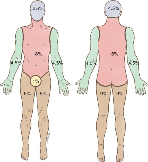
Intact skin provides protection against the environment for all underlying tissues and organs. It aids in heat regulation, prevents water loss, and is the major barrier against bacterial invasion. The tissue injury resulting from a burn disrupts this normal protective function, resulting in local and systemic effects (Box 13-2). Burn patients are therefore some of the most acutely ill patients brought to the OR. The greater the degree of injury to the skin, expressed in percentage of total body surface area (BSA) and depth of burn, the more severe the injury. One method of measuring BSA in adults is by use of the rule of nines (Song et al, 2007) (Figure 13-11).


BOX 13-2


Pathophysiology of Burn Injuries


Thermal and chemical injuries disrupt the normal protective function of the skin, causing local and systemic effects. The extent of these effects depends on the type, duration, and intensity of exposure to the causative agent. With electrical burns, heat is generated as the electrical current passes through body tissues, causing thermal burns along the path taken by the current. Local damage is marked by histamine release and severe vasoconstriction, followed in a few hours by vasodilation and increased capillary permeability, which allows plasma to escape into the wound. Damaged cells swell and platelets and leukocytes aggregate, causing thrombotic ischemia and escalating tissue damage. Systemic effects, which are caused by vascular changes and tissue loss, include hypovolemia, hyperventilation, increased blood viscosity, and suppression of the immune system. The severity of the burn determines the extent of local and systemic effects. Severity is judged by the depth of the burn and the quantity of tissue involved. The depth of the burn is classified by degree. First-degree (superficial) burns affect the epidermis only; second-degree burns (split thickness) affect the epidermis and dermis; third-degree burns (full thickness) affect all skin layers and extend to subcutaneous tissue, muscle, and nerves; fourth-degree burns involve all skin layers, plus bone. The percentage of body surface area (BSA) system of the American Burn Association classifies quantity as follows:


From Langford RW, Thompson JD: Mosby’s handbook of diseases, ed 3, St Louis, 2005, Mosby.


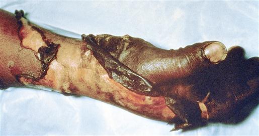
Partial-thickness (first- and second-degree) burns heal by regeneration of skin from dermal elements that remain intact. First-degree burns involve the epidermis, which appears pink or red; sunburn is usually a first-degree burn. Second-degree burns, also called partial-thickness burns, involve the epidermis and some of the dermis. Full-thickness (third-degree) burns (Figure 13-12) involve the epidermis, the entire dermis, and the subcutaneous tissues; they require skin grafting to heal because no dermal elements remain intact. Both partial- and full-thickness burns may require debridement of necrotic tissue (eschar) before healing can occur by skin regeneration or grafting. An allograft may be used to cover the burned area during the initial healing process. However, the allograft must be carefully tested for immunodeficiency diseases. A xenograft (e.g., pig skin) may also be used for covering the burned area.


Procedural Considerations


The essentials of skin grafting are discussed under Skin and Tissue Grafting. This section therefore deals only with the procedure for debridement of burn wounds.


A basic plastic instrument set is required, plus a knife dermatome, an ESU, topical thrombin solution, a pneumatic tourniquet for isolated extremity burns, and a topical antimicrobial agent of choice.


Because patients who have sustained burns are vulnerable to hypothermia from the loss of body surface area (BSA), the temperature and humidity in the OR are increased and exposure is limited only to the areas related to the planned surgical event. Anesthesia is often induced while the patient is on the burn unit bed; transfer to the OR bed is done carefully and gently, with attention to maintaining the airway. Most burn patients arrive in the OR with dressings covering their wounds. The dressings are removed after the patient has been anesthetized to minimize pain and loss of body heat through the open burn wounds. Throughout the procedure, the temperature in the OR is constantly monitored to prevent hypothermia. The OR team caring for burn patients coordinates activities to prevent any delays in obtaining required equipment or supplies. A variety of topical agents are used to dress wounds.


EVIDENCE FOR PRACTICE


Melanoma Awareness, Prevention, and Detection


It is estimated that more than 68,720 men and women will be diagnosed with melanoma in 2009, according to the American Cancer Society. In the most recent time period, rapid increases have occurred among young white women (3.8% annual increase since 1995 in those aged 15 to 34 years) and older white men (8.8% annual increase since 2003 in those 65 and older). Although the exact cause of developing a melanoma is not known, certain risk factors have been identified:


PREVENTION


Limit UV radiation exposure:


KNOW YOUR A-B-C-Ds


Modified from American Cancer Society (ACS): Cancer facts and figures 2009, Atlanta, 2009, The Society.


Operative Procedure


Although skin grafting may be done at the time of wound debridement, it is usually performed several days later, particularly in burns that are extensive.


EXCISIONAL DEBRIDEMENT


Excisional debridement is the act of removing dead or devitalized tissue to promote healing. Plastic surgeons use debridement in conjunction with treatment of injuries, trauma, and infection.


TREATMENT OF PRESSURE ULCERS


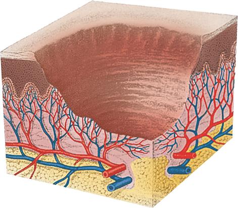
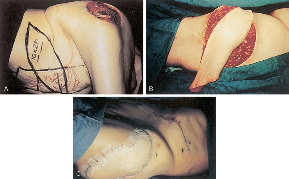
Pressure ulcers result from prolonged compression of soft tissues overlying bony prominences (Figure 13-13). However, whether excessive pressure is sufficient to create an ulcer depends on the intensity and duration of the pressure as well as on tissue tolerance. Factors that contribute to pressure ulcer development are immobility, sensory and motor deficits, reduced circulation, anemia, edema, infection, moisture, shearing force, friction, and nutritional debilitation (Cuzzell and Workman, 2010). The most common sites of pressure ulcers are the sacrum, the ischium, the trochanter, the malleolus, and the heel; these are called decubitus ulcers. These ulcers are different from chronic ulcers such as vascular, diabetic, and neurogenic ulcers. Surgical interventions for pressure ulcers are usually based on ulcer staging (also referred to as grading). In stage I the ulcer involves the epidermis and has soft tissue swelling that is irregular and ill-defined; heat and erythema at the ulcer site are characteristic. A stage II ulcer involves the epidermis and dermis but not the subcutaneous fat. Stage III ulcers show full-thickness skin loss with injury to underlying tissue layers and may contain necrotic material. Thorough excisional debridement is performed, and IV antibiotic therapy is instituted. Although debrided stage III ulcers often heal on their own, surgical excision and closure may be done to prevent a lengthy spontaneous closure, which may result in a weak, unstable scar with resultant recurrence. Stage IV ulcers are the deepest, requiring more radical excisional debridement. Adequate soft tissue cover may be obtained by either split-thickness or full-thickness skin grafting or tissue flaps (Figure 13-14). Tissue expansion may be used when there is not enough tissue adjacent to the ulcer site to provide flap coverage.


The use of a CO2 laser minimizes blood loss and possibly reduces infection rates in the presence of gross contamination. Although many techniques and flaps are surgical options, basic principles apply to all pressure ulcer closure procedures. The following procedure is for an adjacent flap.


Procedural Considerations


A basic plastic instrument set is required, as well as assorted sizes of osteotomes (straight and curved), a mallet, the Gigli saw and handle, assorted curettes, a Key periosteal elevator, a duckbill rongeur, bone wax, the dermatome of choice, the ESU, a sterile marking pen, and a closed-wound drainage system. The patient is positioned and draped so that the pressure ulcer, adjacent flap donor site, and skin graft donor site are well exposed.


Operative Procedure


SURGICAL PHARMACOLOGY


Topical Medications Used in Burn Therapy


Agent Indication Implications
Petroleum-based antimicrobials (bacitracin, Neosporin, polymyxin B) Partial-thickness burns
Silver sulfadiazine (Silvadene)

Mafenide acetate (Sulfamylon)

Stay updated, free articles. Join our Telegram channel

Dec 9, 2016 | Posted by in GENERAL & FAMILY MEDICINE | Comments Off on 13. Plastic and Reconstructive Surgery

Full access? Get Clinical Tree

Get Clinical Tree app for offline access