13 CASE 13
A 22-year-old female college student comes to the university clinic complaining of palpitations.
PATHOPHYSIOLOGY OF KEY SYMPTOMS
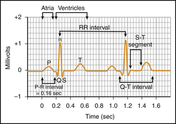
The electrocardiogram provides a record of the flow of current that accompanies depolarization of the heart. The normal electrocardiogram consists of a P wave, representing atrial depolarization, a QRS complex representing ventricular depolarization, and a T wave representing ventricular repolarization. This sequence is termed “normal sinus rhythm.” Spread of the depolarization through the AV node is contained within the PR interval (Fig. 13-1).
< div class='tao-gold-member'>
Only gold members can continue reading. Log In or Register to continue
Stay updated, free articles. Join our Telegram channel

Full access? Get Clinical Tree


