11

11 CASE 11


A 4-day-old female infant is seen by the cardiologist, having been referred because of suspicion of a patent ductus arteriosus.


The infant was delivered by cesarean section at 32 weeks’ gestational age because of preeclampsia in the mother. The APGAR score was 7 at 1 minute and improved to 9 at 5 minutes. The pediatrician noted a machine-like murmur extending through both the systole and diastole.


PATHOPHYSIOLOGY OF KEY SYMPTOMS


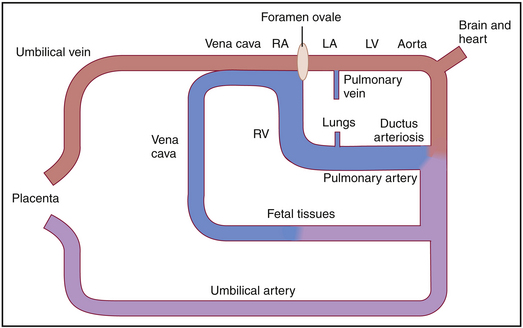
In utero, the ductus arteriosus allows the flow of blood from the pulmonary artery into the aorta, so that 90% of the right ventricular output bypasses the lungs. Blood flowing through the ductus arteriosus joins with the blood pumped by the left ventricle into the aorta to provide blood flow to most of the body. Vessels that originate from the aorta before the juncture with the ductus arteriosus carry relatively well oxygenated blood to the brain and heart (Fig. 11-1).


At birth, the inflation of the lungs reduces pulmonary vascular resistance and the constriction of the umbilical artery increases systemic vascular resistance. Consequently, aortic pressure becomes higher than pulmonary artery pressure and oxygenated blood flows from the aorta into the pulmonary artery. The presence of oxygenated blood in the ductus arteriosus normally inhibits the production of the vasodilator prostaglandins, and the ductus arteriosus closes. Closure of the ductus arteriosus, along with closure of the foramen ovale, effectively isolates the pulmonary circulation from the systemic circulation. Remnants of the ductus arteriosus persist throughout life and are identified as the ligamentum arteriosus connecting the pulmonary artery and the aorta.


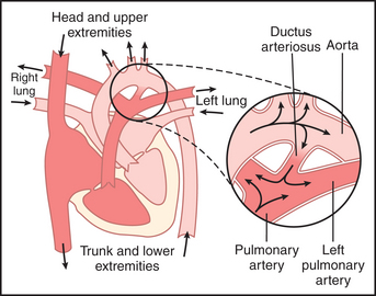
When the ductus arteriosus remains patent, or “open,” blood flow from the aorta into the pulmonary artery persists after birth. There may not be any apparent symptoms, because this congenital defect does not impair oxygenation of blood pumped by the left ventricle. Instead, blood flowing through the ductus arteriosus actually passes through the lungs for a second time before being pumped into the systemic circulation (Fig. 11-2).


< div class='tao-gold-member'>

Stay updated, free articles. Join our Telegram channel

Jul 4, 2016 | Posted by in PHYSIOLOGY | Comments Off on 11

Full access? Get Clinical Tree

Get Clinical Tree app for offline access