CASE 11
A 65-year-old retired plasterer presented to his physician with complaints of chest pain and difficulty breathing. He also complained of weakness. The physical examination was unremarkable. Chest radiographs indicated a large diffuse mass in the right pleural cavity. A thoracotomy was performed to obtain a biopsy. The pathologist’s report concluded that the patient had malignant mesothelioma.
WHAT ARE THE SURFACE MARKINGS OF THE INFERIOR LIMITS OF THE PARIETAL PLEURA, VISCERAL PLEURA, AND LUNGS AFTER EXPIRATION?
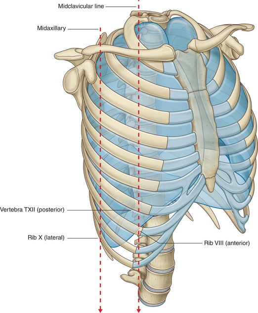
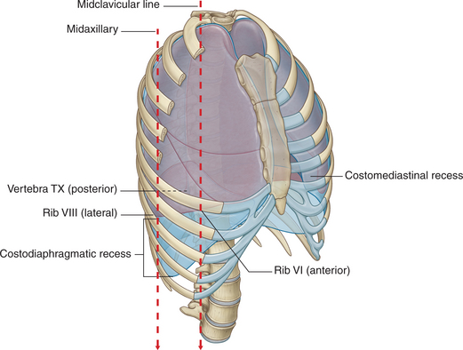
The inferior surface markings of these structures are based on three vertical lines. These are midclavicular, midaxillary, and vertebral. The structures that define the inferior limits are the ribs. Moving from the midclavicular to vertebral lines, the inferior limits of the parietal pleura are represented by ribs 8, 10, and 12 (Fig. 2-26). Because the visceral pleura is adhered to the surface of the lung, both these structures share the same limits. Again, proceeding from the midclavicular to the vertebral lines, the inferior limits are ribs 6, 8, and 10 (Fig. 2-27).

FIGURE 2-26 Topography of the parietal pleura to the ribs.
(Drake R, Vogl W and Mitchell A: Gray’s Anatomy for Students. Churchill Livingstone, 2004. Fig. 3-37.)


